
Search Count: 166
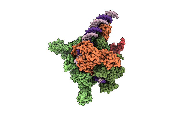 |
Cryo-Em Structure Of Vibrio Cholerae Rna Polymerase Holoenzyme Bound To An Ompu Promoter Dna Fragment
Organism: Vibrio cholerae o395
Method: ELECTRON MICROSCOPY Release Date: 2025-12-17 Classification: TRANSCRIPTION Ligands: MG, ZN |
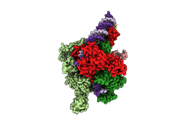 |
Cryo-Em Structure Of Vibrio Cholerae Rna Polymerase Holoenzyme Bound To An Ompu Promoter Dna Fragment And 5-Mer Rna
Organism: Vibrio cholerae o395
Method: ELECTRON MICROSCOPY Release Date: 2025-12-17 Classification: TRANSCRIPTION Ligands: ZN, MG |
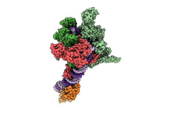 |
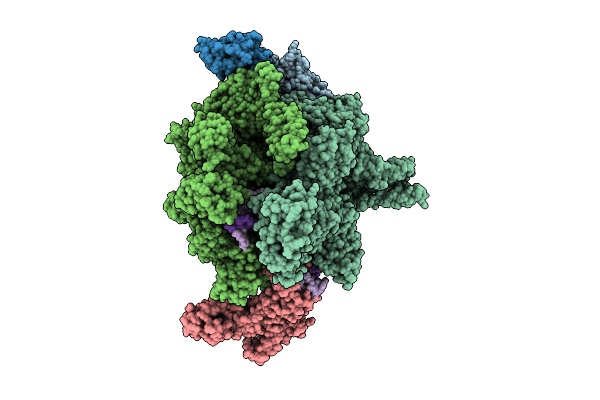
Cryo-Em Structure Of Vibrio Cholerae Rna Polymerase Transcription Activation Complex With Toxr Transcription Factor And Ompu Promoter Dna
Organism: Vibrio cholerae o395, Vibrio cholerae o1 biovar el tor str. n16961
Method: ELECTRON MICROSCOPY Release Date: 2025-12-17 Classification: TRANSCRIPTION Ligands: ZN, MG |
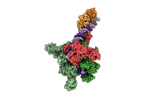 |
Cryo-Em Structure Of Vibrio Cholerae Rna Polymerase Transcription Activation Complex With Tcpp Transcription Factor And A Toxt Promoter Dna Fragment
Organism: Vibrio cholerae o395
Method: ELECTRON MICROSCOPY Release Date: 2025-12-17 Classification: TRANSCRIPTION Ligands: ZN, MG |
 |
Cryo-Em Structure Of Vibrio Cholerae Rna Polymerase Transcription Activation Complex With Toxr And Tcpp Transcription Factors And A Toxt Promoter Dna Fragment
Organism: Vibrio cholerae o395, Vibrio cholerae o1 biovar el tor str. n16961
Method: ELECTRON MICROSCOPY Release Date: 2025-12-17 Classification: TRANSCRIPTION Ligands: MG, ZN |
 |
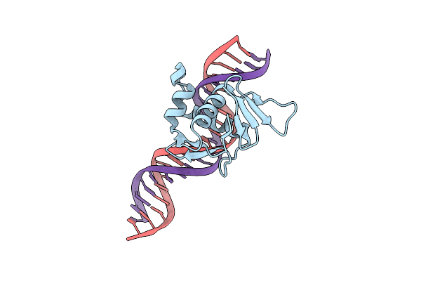
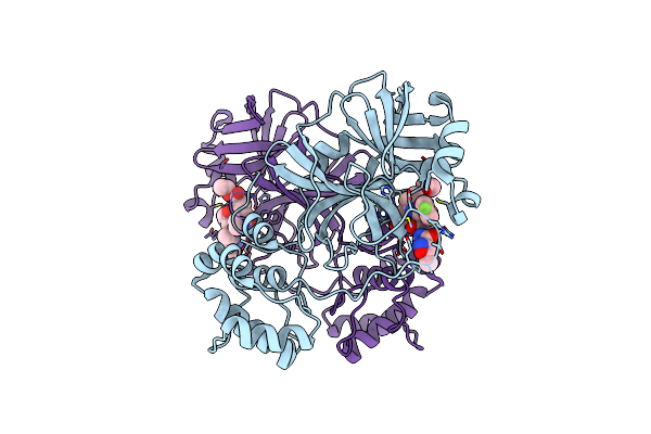
Organism: Vibrio cholerae
Method: X-RAY DIFFRACTION Resolution:1.75 Å Release Date: 2023-08-09 Classification: DNA BINDING PROTEIN Ligands: CD, NH4 |
 |
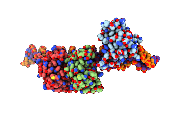
Organism: Vibrio cholerae
Method: X-RAY DIFFRACTION Resolution:2.07 Å Release Date: 2023-08-09 Classification: DNA BINDING PROTEIN |
 |
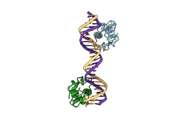
Organism: Vibrio cholerae
Method: X-RAY DIFFRACTION Resolution:2.64 Å Release Date: 2023-08-09 Classification: DNA BINDING PROTEIN |
 |
Organism: Vibrio cholerae
Method: X-RAY DIFFRACTION Resolution:3.25 Å Release Date: 2023-08-09 Classification: DNA BINDING PROTEIN |
 |
Organism: Streptococcus agalactiae
Method: X-RAY DIFFRACTION Resolution:1.50 Å Release Date: 2023-02-08 Classification: REPLICATION Ligands: GOL, MN, CL |
 |
Organism: Streptococcus agalactiae
Method: X-RAY DIFFRACTION Resolution:3.00 Å Release Date: 2023-02-08 Classification: REPLICATION Ligands: MN |
 |
Organism: Streptococcus agalactiae
Method: X-RAY DIFFRACTION Resolution:2.77 Å Release Date: 2023-02-08 Classification: REPLICATION Ligands: PO4, NA |
 |
Organism: Severe acute respiratory syndrome coronavirus 2
Method: X-RAY DIFFRACTION Resolution:2.26 Å Release Date: 2022-12-28 Classification: VIRAL PROTEIN Ligands: XNV |
 |
Organism: Severe acute respiratory syndrome coronavirus
Method: X-RAY DIFFRACTION Resolution:2.53 Å Release Date: 2022-12-28 Classification: VIRAL PROTEIN Ligands: XNV |
 |
Organism: Severe acute respiratory syndrome coronavirus 2
Method: X-RAY DIFFRACTION Resolution:2.26 Å Release Date: 2021-07-21 Classification: VIRAL PROTEIN Ligands: AG7 |
 |
Crystal Structure Of T7 Bacteriophage Portal Protein, 13Mer, Closed Valve - P212121
Organism: Enterobacteria phage t7
Method: X-RAY DIFFRACTION Resolution:3.74 Å Release Date: 2020-12-16 Classification: VIRAL PROTEIN |
 |
Organism: Epstein-barr virus (strain gd1)
Method: ELECTRON MICROSCOPY Release Date: 2019-09-18 Classification: VIRAL PROTEIN |
 |
Organism: Epstein-barr virus (strain gd1)
Method: ELECTRON MICROSCOPY Release Date: 2019-09-18 Classification: VIRAL PROTEIN |
 |
Organism: Enterobacteria phage t7
Method: X-RAY DIFFRACTION Resolution:3.40 Å Release Date: 2019-09-04 Classification: VIRAL PROTEIN |
 |
Organism: Enterobacteria phage t7
Method: X-RAY DIFFRACTION Resolution:3.60 Å Release Date: 2019-09-04 Classification: VIRAL PROTEIN |