
Search Count: 55
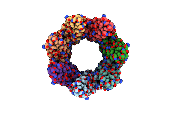 |
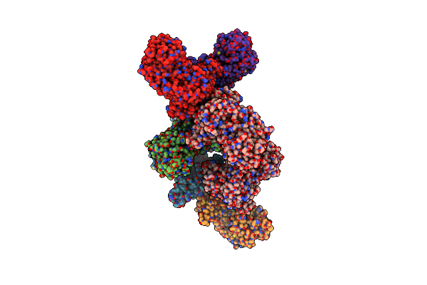
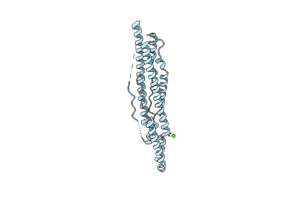
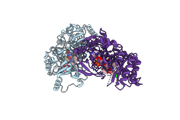
Cryoem Structure Of The Ring-Shaped Virulence Factor Espb From Mycobacterium Tuberculosis
Organism: Mycobacterium tuberculosis (strain atcc 25618 / h37rv)
Method: ELECTRON MICROSCOPY Release Date: 2020-06-17 Classification: TRANSPORT PROTEIN |
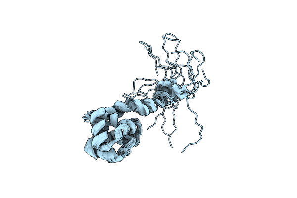 |
Structural And Dna Binding Properties Of Mycobacterial Integration Host Factor Mihf
Organism: Mycobacterium tuberculosis
Method: SOLUTION NMR Release Date: 2019-12-25 Classification: DNA BINDING PROTEIN |
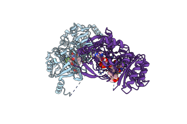 |
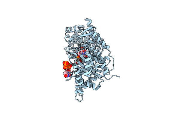
Crystal Structure Of M. Tuberculosis Dpre1 In Complex With Spbtz169 (Sulfonylpbtz)
Organism: Mycobacterium tuberculosis (strain atcc 25618 / h37rv)
Method: X-RAY DIFFRACTION Release Date: 2018-08-01 Classification: OXIDOREDUCTASE Ligands: FAD, EQ8 |
 |
Organism: Mycobacterium thermoresistibile atcc 19527
Method: X-RAY DIFFRACTION Resolution:1.60 Å Release Date: 2016-10-19 Classification: OXIDOREDUCTASE Ligands: 6G1, IMP |
 |
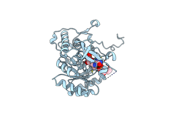
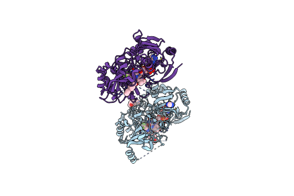
Crystal Structure Of M. Tuberculosis Dpre1 In Complex With The Nitro-Benzothiazole 6A
Organism: Mycobacterium tuberculosis
Method: X-RAY DIFFRACTION Resolution:3.00 Å Release Date: 2015-10-28 Classification: OXIDOREDUCTASE/OXIDOREDUCTASE INHIBITOR Ligands: FAD, O95 |
 |
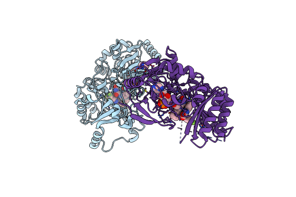
Organism: Mycobacterium tuberculosis (strain atcc 25618 / h37rv)
Method: X-RAY DIFFRACTION Resolution:3.52 Å Release Date: 2015-07-01 Classification: LIGASE Ligands: CA |
 |
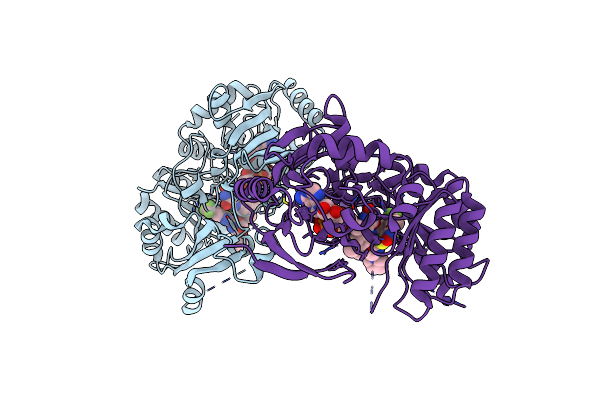
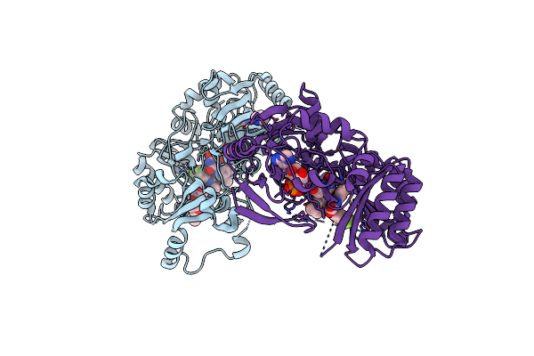
Crystal Structure Of The M. Tuberculosis Ctp Synthase Pyrg In Complex With Two Utp Molecules
Organism: Mycobacterium tuberculosis (strain atcc 25618 / h37rv)
Method: X-RAY DIFFRACTION Resolution:1.99 Å Release Date: 2015-07-01 Classification: LIGASE Ligands: UTP, MG, GOL |
 |
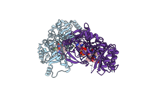
Crystal Structure Of The M. Tuberculosis Ctp Synthase Pyrg In Complex With Utp, Amp-Pcp And Oxonorleucine
Organism: Mycobacterium tuberculosis (strain atcc 25618 / h37rv)
Method: X-RAY DIFFRACTION Resolution:3.49 Å Release Date: 2015-07-01 Classification: LIGASE Ligands: ONL, ACP, UTP, MG |
 |
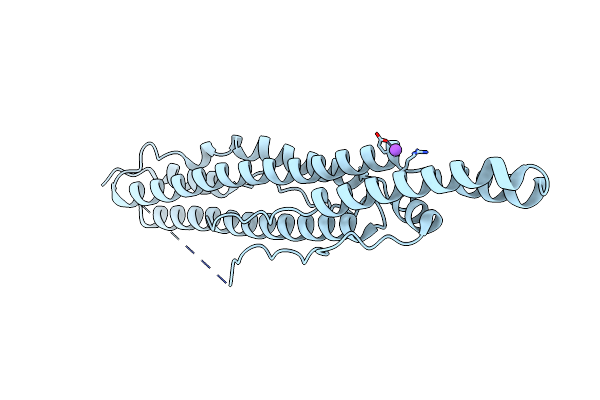
Structure Of Pe-Ppe Domains Of Esx-1 Secreted Protein Espb, C2221 In Presence Of Ca
Organism: Mycobacterium tuberculosis
Method: X-RAY DIFFRACTION Resolution:1.82 Å Release Date: 2015-02-18 Classification: PROTEIN TRANSPORT Ligands: CA |
 |
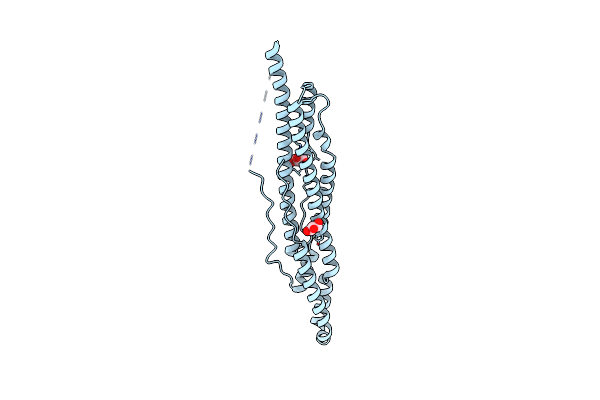
Organism: Mycobacterium tuberculosis
Method: X-RAY DIFFRACTION Resolution:2.14 Å Release Date: 2015-02-18 Classification: PROTEIN TRANSPORT Ligands: CL, NA |
 |
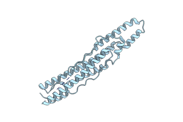
Organism: Mycobacterium tuberculosis
Method: X-RAY DIFFRACTION Resolution:1.50 Å Release Date: 2015-02-18 Classification: PROTEIN TRANSPORT Ligands: CL, GOL |
 |
Organism: Mycobacterium tuberculosis
Method: X-RAY DIFFRACTION Resolution:3.04 Å Release Date: 2015-02-18 Classification: PROTEIN TRANSPORT |
 |
Organism: Corynebacterium glutamicum, Synthetic construct
Method: X-RAY DIFFRACTION Resolution:1.82 Å Release Date: 2014-12-17 Classification: TRANSCRIPTION Ligands: CMP, GOL |
 |
Crystal Structure Of The M. Tuberculosis Sulfate Ester Dioxygenase Rv3406 In Complex With Iron.
Organism: Mycobacterium tuberculosis
Method: X-RAY DIFFRACTION Resolution:2.00 Å Release Date: 2014-12-10 Classification: OXIDOREDUCTASE Ligands: NO3, FE |
 |
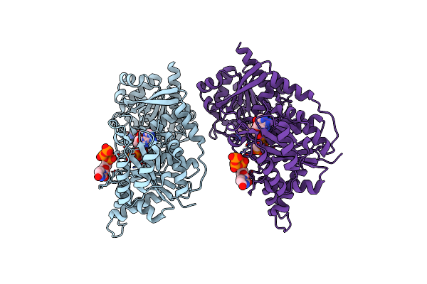
Crystal Structure Of M. Tuberculosis Dpre1 In Complex With The Non-Covalent Inhibitor Qn127
Organism: Mycobacterium tuberculosis
Method: X-RAY DIFFRACTION Resolution:1.95 Å Release Date: 2014-12-10 Classification: OXIDOREDUCTASE/OXIDOREDUCTASE INHIBITOR Ligands: FAD, Y22, IMD, 2J3 |
 |
Crystal Structure Of M. Tuberculosis Dpre1 In Complex With The Non-Covalent Inhibitor Ty38C
Organism: Mycobacterium tuberculosis
Method: X-RAY DIFFRACTION Resolution:2.49 Å Release Date: 2014-12-10 Classification: OXIDOREDUCTASE/OXIDOREDUCTASE INHIBITOR Ligands: FAD, 38C |
 |
Crystal Structure Of M. Tuberculosis Dpre1 In Complex With The Non-Covalent Inhibitor Ty36C
Organism: Mycobacterium tuberculosis
Method: X-RAY DIFFRACTION Resolution:2.02 Å Release Date: 2014-12-10 Classification: OXIDOREDUCTASE/OXIDOREDUCTASE INHIBITOR Ligands: FAD, 36C, IMD |
 |
Crystal Structure Of M. Tuberculosis Dpre1 In Complex With The Non-Covalent Inhibitor Qn114
Organism: Mycobacterium tuberculosis
Method: X-RAY DIFFRACTION Resolution:2.09 Å Release Date: 2014-12-10 Classification: OXIDOREDUCTASE/OXIDOREDUCTASE INHIBITOR Ligands: FAD, R58, 2J3, IMD |
 |
Crystal Structure Of M. Tuberculosis Dpre1 In Complex With The Non-Covalent Inhibitor Qn118
Organism: Mycobacterium tuberculosis
Method: X-RAY DIFFRACTION Resolution:1.79 Å Release Date: 2014-12-10 Classification: OXIDOREDUCTASE/OXIDOREDUCTASE INHIBITOR Ligands: FAD, R57, IMD |
 |
Crystal Structure Of M. Tuberculosis Dpre1 In Complex With The Non-Covalent Inhibitor Qn127
Organism: Mycobacterium tuberculosis
Method: X-RAY DIFFRACTION Resolution:2.20 Å Release Date: 2014-12-10 Classification: OXIDOREDUCTASE/OXIDOREDUCTASE INHIBITOR Ligands: FAD, RG2, 2J3, IMD |