
Search Count: 21
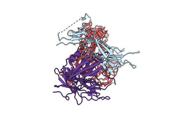 |
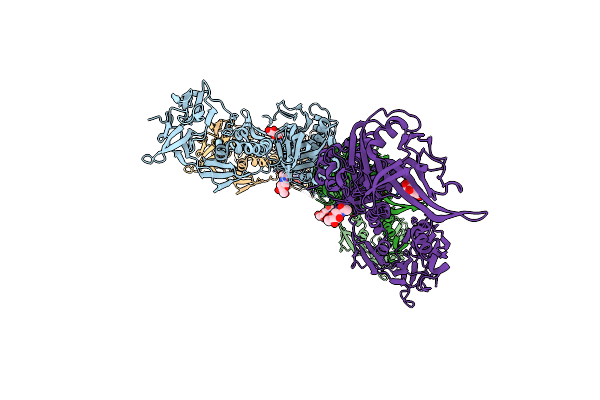
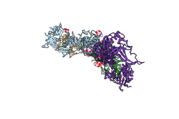
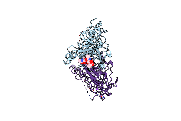
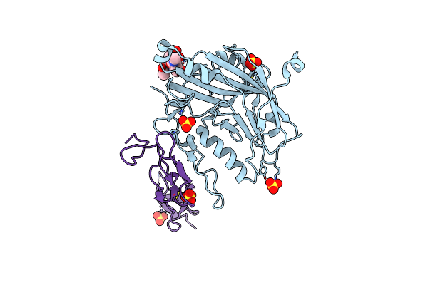
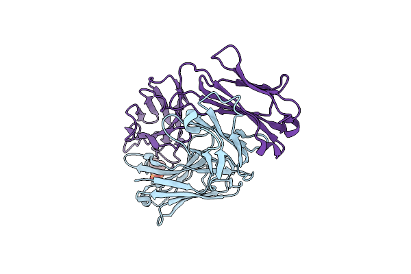
Herpes Simplex Virus 2 Delta28-73 Glycoprotein C Ectodomain In Complex With C3B
Organism: Human alphaherpesvirus 2, Homo sapiens
Method: ELECTRON MICROSCOPY Release Date: 2025-12-10 Classification: VIRAL PROTEIN |
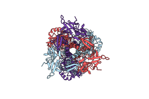 |
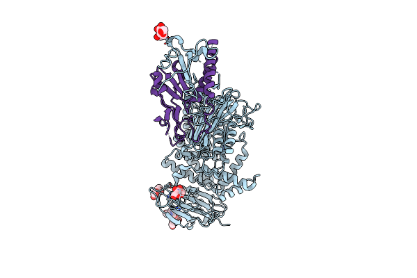
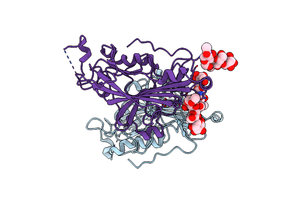
Structure Of Herpesvirus Fusion Glycoprotein B-Bilayer Complex Revealing The Protein-Membrane And Lateral Protein-Protein Interaction
Organism: Human herpesvirus 1
Method: ELECTRON MICROSCOPY Resolution:27.00 Å Release Date: 2013-07-31 Classification: VIRAL PROTEIN |
 |
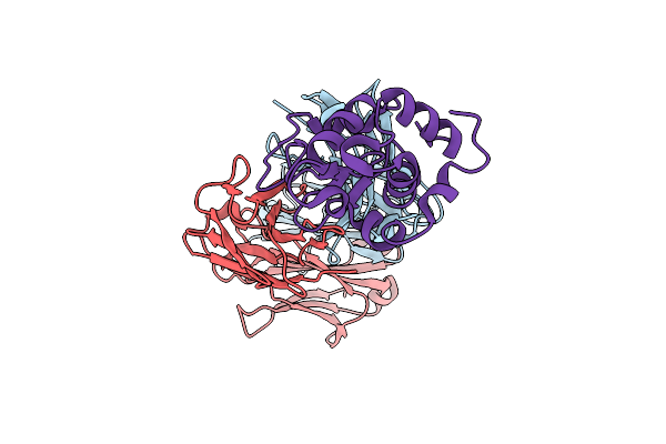
Organism: Human herpesvirus 1, Homo sapiens
Method: X-RAY DIFFRACTION Resolution:4.00 Å Release Date: 2011-10-12 Classification: VIRAL PROTEIN/PROTEIN BINDING Ligands: NAG |
 |
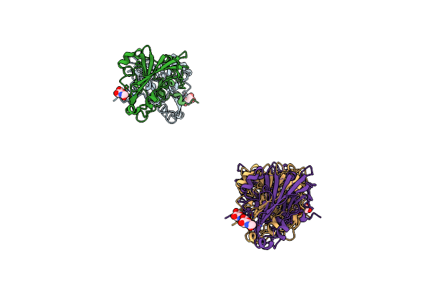
Organism: Human herpesvirus 1
Method: X-RAY DIFFRACTION Resolution:2.76 Å Release Date: 2010-12-01 Classification: VIRAL PROTEIN Ligands: NAG, MRY, NA, CL |
 |
Organism: Human herpesvirus 1
Method: X-RAY DIFFRACTION Resolution:2.26 Å Release Date: 2010-12-01 Classification: VIRAL PROTEIN Ligands: NAG, MRY |
 |
Organism: Human herpesvirus 1
Method: X-RAY DIFFRACTION Resolution:2.88 Å Release Date: 2010-12-01 Classification: VIRAL PROTEIN Ligands: NAG, CL, MRY |
 |
Organism: Human herpesvirus 1
Method: X-RAY DIFFRACTION Resolution:3.00 Å Release Date: 2010-12-01 Classification: VIRAL PROTEIN Ligands: NAG, MRY, NA |
 |
Crystal Structure Of The Conserved Herpesvirus Fusion Regulator Complex Gh-Gl
Organism: Human herpesvirus 2
Method: X-RAY DIFFRACTION Resolution:3.00 Å Release Date: 2010-07-07 Classification: VIRAL PROTEIN Ligands: NAG, XYL |
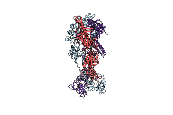 |
Crystal Structure Of The Extracellular Domain Of Glycoprotein B From Herpes Simplex Virus Type I
Organism: Human herpesvirus 1
Method: X-RAY DIFFRACTION Resolution:2.10 Å Release Date: 2006-07-25 Classification: VIRAL PROTEIN |
 |
Structure Of Unliganded Hsv Gd Reveals A Mechanism For Receptor- Mediated Activation Of Virus Entry
Organism: Human herpesvirus 1
Method: X-RAY DIFFRACTION Resolution:2.50 Å Release Date: 2005-12-21 Classification: VIRAL PROTEIN Ligands: CL, ZN, NA |
 |
Structure Of Unliganded Hsv Gd Reveals A Mechanism For Receptor- Mediated Activation Of Virus Entry
Organism: Human herpesvirus 1
Method: X-RAY DIFFRACTION Resolution:2.11 Å Release Date: 2005-11-23 Classification: VIRAL PROTEIN Ligands: ZN, CL |
 |
The Crystal Structure Of The Antibody Fab Hyhel5 Complex With Lysozyme At 1.7A Resolution
Organism: Mus musculus, Gallus gallus
Method: X-RAY DIFFRACTION Resolution:1.70 Å Release Date: 2005-04-26 Classification: IMMUNE SYSTEM |
 |
Organism: Human herpesvirus 1
Method: X-RAY DIFFRACTION Resolution:2.85 Å Release Date: 2003-12-16 Classification: VIRAL PROTEIN Ligands: NAG |
 |
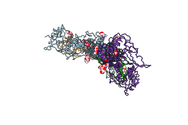
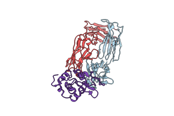
Crystal Structure Of The Herpes Simplex Virus Glycoprotein D Bound To The Cellular Receptor Hvea/Hvem
Organism: Human herpesvirus 1, Homo sapiens
Method: X-RAY DIFFRACTION Resolution:2.65 Å Release Date: 2001-09-26 Classification: VIRAL PROTEIN Ligands: SO4 |
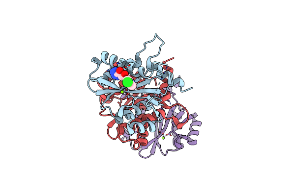 |
Core Domain Of Hiv-1 Integrase Complexed With Mg++ And 1-(5-Chloroindol-3-Yl)-3-Hydroxy-3-(2H-Tetrazol-5-Yl)-Propenone
Organism: Human immunodeficiency virus 1
Method: X-RAY DIFFRACTION Resolution:2.10 Å Release Date: 1999-11-17 Classification: TRANSFERASE Ligands: MG, 100 |
 |
Organism: Homo sapiens
Method: X-RAY DIFFRACTION, SOLUTION NMR Resolution:2.00 Å Release Date: 1996-08-17 Classification: CYTOKINE |
 |
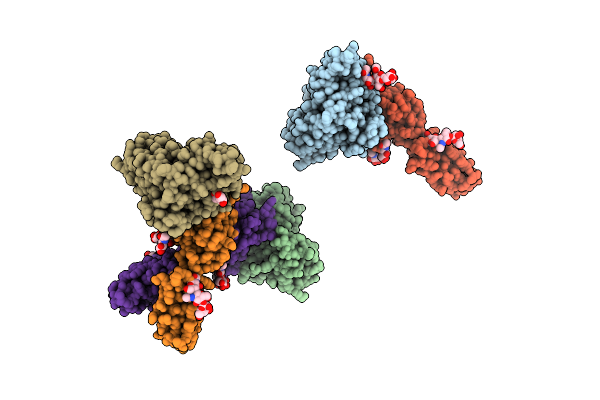
Structure Of An Antibody-Antigen Complex. Crystal Structure Of The Hy/Hel-10 Fab-Lysozyme Complex
Organism: Mus musculus, Gallus gallus
Method: X-RAY DIFFRACTION Resolution:3.00 Å Release Date: 1989-07-12 Classification: COMPLEX(ANTIBODY-ANTIGEN) Ligands: HOH |
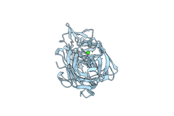 |
Structure And Refinement At 1.8 Angstroms Resolution Of The Aspartic Proteinase From Rhizopus Chinensis
Organism: Rhizopus microsporus var. chinensis
Method: X-RAY DIFFRACTION Resolution:1.80 Å Release Date: 1987-07-16 Classification: HYDROLASE (ASPARTIC PROTEINASE) Ligands: CA |
 |
Phosphocholine Binding Immunoglobulin Fab Mc/Pc603. An X-Ray Diffraction Study At 2.7 Angstroms
Organism: Mus musculus
Method: X-RAY DIFFRACTION Resolution:2.70 Å Release Date: 1985-01-02 Classification: IMMUNOGLOBULIN Ligands: SO4 |
 |
Refined Crystal Structure Of The Mc/Pc603 Fab-Phosphocholine Complex At 3.1 Angstroms Resolution
Organism: Mus musculus
Method: X-RAY DIFFRACTION Resolution:3.10 Å Release Date: 1985-01-02 Classification: IMMUNE SYSTEM Ligands: PC |