
Search Count: 54
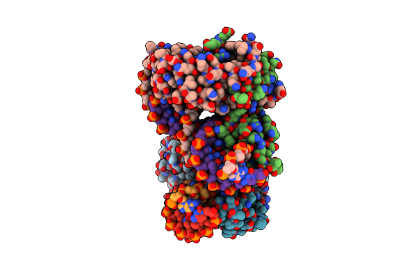 |
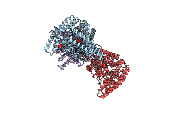
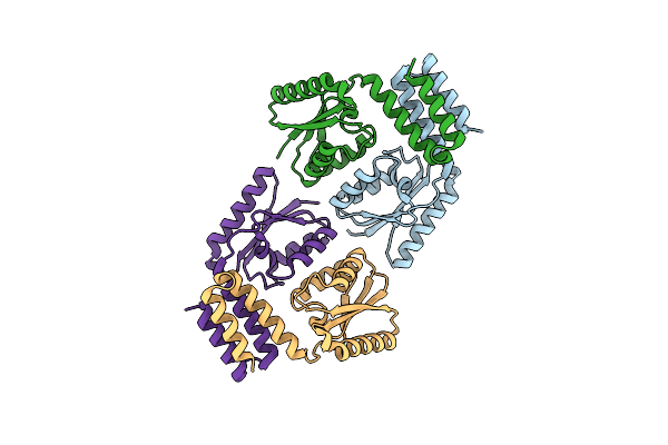
Crystal Structure Of Putative Marr Family Transcriptional Regulator Hcar From Acinetobacter Sp. Adp Complexed With 24Mer Dna.
Organism: Acinetobacter baylyi (strain atcc 33305 / bd413 / adp1), Acinetobacter sp. adp1
Method: X-RAY DIFFRACTION Resolution:3.00 Å Release Date: 2015-10-14 Classification: TRANSCRIPTION |
 |
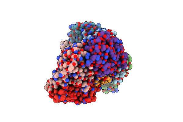
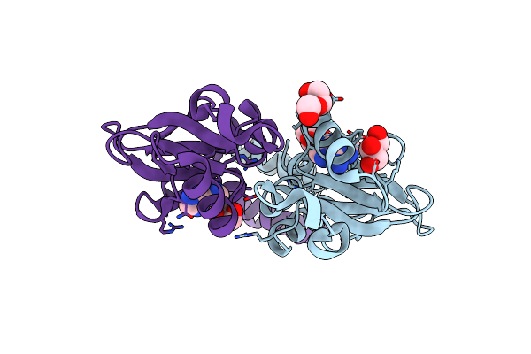
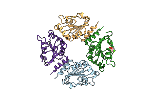
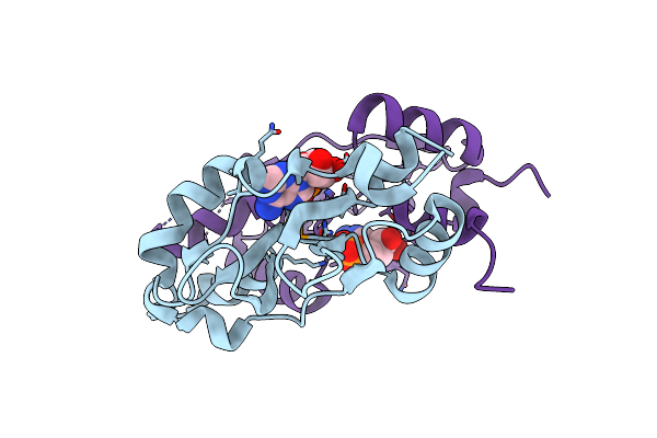
Crystal Structure Of Putative Marr Family Transcriptional Regulator Hcar From Acinetobacter Sp. Adp
Organism: Acinetobacter sp. adp1
Method: X-RAY DIFFRACTION Resolution:2.30 Å Release Date: 2015-03-04 Classification: TRANSCRIPTION Ligands: HC4, SO4, GOL |
 |
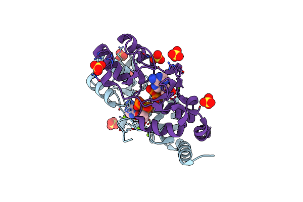
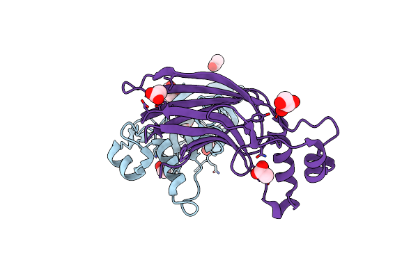
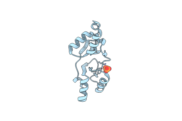
Crystal Structure Of Putative Marr Family Transcriptional Regulator Hcar From Acinetobacter Sp. Adp Complexed With Vanilin
Organism: Acinetobacter sp. adp1
Method: X-RAY DIFFRACTION Resolution:2.30 Å Release Date: 2015-03-04 Classification: TRANSCRIPTION Ligands: V55, SO4, GOL |
 |
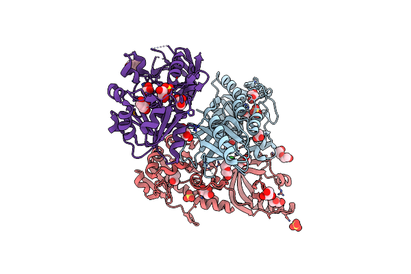
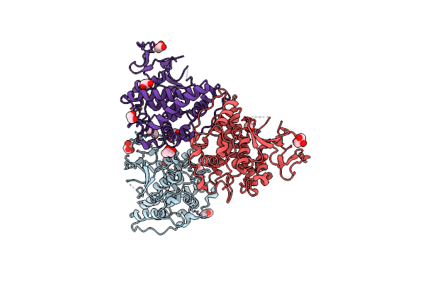
Crystal Structure Of Putative Marr Family Transcriptional Regulator Hcar From Acinetobacter Sp. Adp Complexed With Ferulic Acid
Organism: Acinetobacter sp. adp1
Method: X-RAY DIFFRACTION Resolution:1.89 Å Release Date: 2015-03-04 Classification: TRANSCRIPTION Ligands: FER, CL, GOL, NA |
 |
Crystal Structure Of Putative Marr Family Transcriptional Regulator Hcar From Acinetobacter Sp. Adp Complexed With 3,4-Dihydroxy Bezoic Acid
Organism: Acinetobacter sp. adp1
Method: X-RAY DIFFRACTION Resolution:2.10 Å Release Date: 2015-03-04 Classification: TRANSCRIPTION Ligands: DHB, EDO, CL |
 |
Organism: Lactobacillus plantarum
Method: X-RAY DIFFRACTION Resolution:1.90 Å Release Date: 2011-06-08 Classification: CHAPERONE Ligands: ATP, CA, GOL, ACT |
 |
The Crystal Structure Of A Domain From A Possible Membrane Protein Of Bordetella Parapertussis
Organism: Bordetella parapertussis
Method: X-RAY DIFFRACTION Resolution:1.80 Å Release Date: 2010-10-06 Classification: MEMBRANE PROTEIN Ligands: MG, ADP, SO4 |
 |
The Crystal Structure Of A Hemolysin-Like Protein Containing Cbs Domain Of Oenococcus Oeni Psu
Organism: Oenococcus oeni
Method: X-RAY DIFFRACTION Resolution:2.20 Å Release Date: 2010-10-06 Classification: structural genomics, unknown function |
 |
Crystal Structure Of Macrolide-Efflux Protein Sp_1110 From Streptococcus Pneumoniae
Organism: Streptococcus pneumoniae
Method: X-RAY DIFFRACTION Resolution:2.49 Å Release Date: 2010-09-29 Classification: TRANSFERASE Ligands: SO4, GOL, PEG, ACY, CL |
 |
The Crystal Structure Of The Putative Lipoprotein From Colwellia Psychrerythraea
Organism: Colwellia psychrerythraea
Method: X-RAY DIFFRACTION Resolution:1.70 Å Release Date: 2010-09-22 Classification: LIPID BINDING PROTEIN Ligands: SO4, TRS, GOL |
 |
The Crystal Structure Of Functionally Unknown Conserved Protein Domain From Neisseria Meningitidis Mc58
Organism: Neisseria meningitidis serogroup b
Method: X-RAY DIFFRACTION Resolution:1.99 Å Release Date: 2010-09-22 Classification: Structural genomics, Unknown function Ligands: ADN, GOL, PEG |
 |
Organism: Sulfurospirillum deleyianum
Method: X-RAY DIFFRACTION Resolution:2.00 Å Release Date: 2010-08-18 Classification: structural genomics, unknown function Ligands: ACY, GOL |
 |
The Structure Of A Putative Glutathione S-Transferase From Corynebacterium Glutamicum
Organism: Corynebacterium glutamicum
Method: X-RAY DIFFRACTION Resolution:2.10 Å Release Date: 2010-04-14 Classification: TRANSFERASE Ligands: GOL, EDO |
 |
The Crystal Structure Of A Thiol-Disulfide Isomerase From Corynebacterium Glutamicum To 2.2A
Organism: Corynebacterium glutamicum
Method: X-RAY DIFFRACTION Resolution:2.20 Å Release Date: 2010-03-16 Classification: ISOMERASE Ligands: CL, CA, ACT |
 |
The Crystal Structure Of The Putative Cell Surface Hydrolase From Lactobacillus Plantarum Wcfs1
Organism: Lactobacillus plantarum
Method: X-RAY DIFFRACTION Resolution:2.00 Å Release Date: 2010-03-16 Classification: HYDROLASE |
 |
Crystal Structure Of Cbs Domain Of A Putative Transporter From Clostridium Difficile 630
Organism: Clostridium difficile 630
Method: X-RAY DIFFRACTION Resolution:2.40 Å Release Date: 2010-03-09 Classification: MEMBRANE PROTEIN Ligands: PO4 |
 |
The Crystal Structure Of A Secreted Thiol-Disulfide Isomerase From Corynebacterium Glutamicum To 1.75A
Organism: Corynebacterium glutamicum
Method: X-RAY DIFFRACTION Resolution:1.75 Å Release Date: 2010-03-02 Classification: ISOMERASE Ligands: CA |
 |
Crystal Structure Of Putative Dna Binding Protein From Methanocaldococcus Jannaschii.
Organism: Methanocaldococcus jannaschii
Method: X-RAY DIFFRACTION Resolution:2.20 Å Release Date: 2010-02-02 Classification: DNA BINDING PROTEIN |
 |
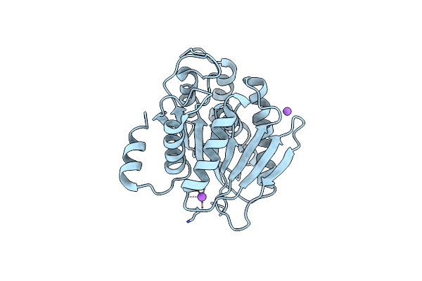
The Crystal Structure Of A Cbs Domain From A Putative Metal Ion Transporter Bound To Amp From Pseudomonas Syringae To 1.55A
Organism: Pseudomonas syringae
Method: X-RAY DIFFRACTION Resolution:1.53 Å Release Date: 2010-01-26 Classification: TRANSPORT PROTEIN Ligands: AMP |
 |
The Crystal Structure Of A Domain Of Phenylacetate-Coenzyme A Ligase From Bacteroides Vulgatus Atcc 8482
Organism: Bacteroides vulgatus
Method: X-RAY DIFFRACTION Resolution:1.43 Å Release Date: 2010-01-19 Classification: LIGASE |