
Search Count: 88
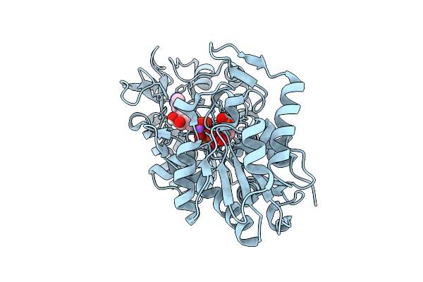 |
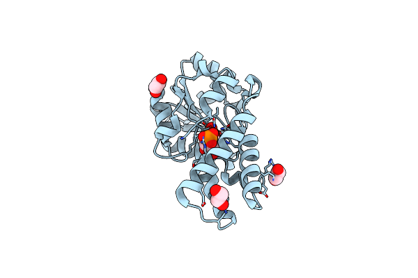
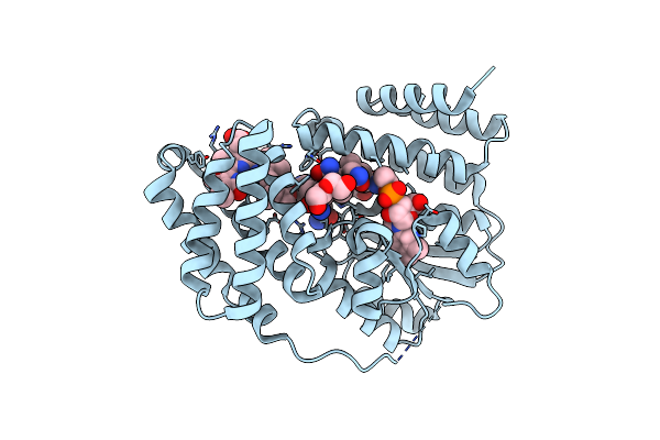
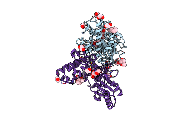
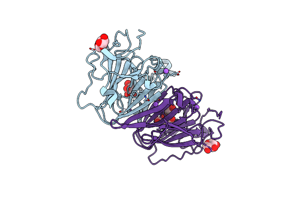
Human Phosphoglycerate Kinase In With Mixture Of Products And Substrates Produced By Cross-Soaking A Tsa Crystal
Organism: Homo sapiens
Method: X-RAY DIFFRACTION Release Date: 2025-10-29 Classification: TRANSFERASE Ligands: ATP, X15, NA, CL |
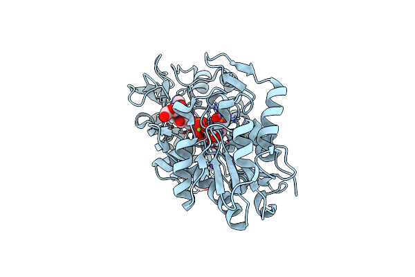 |
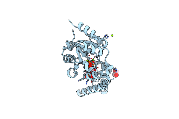
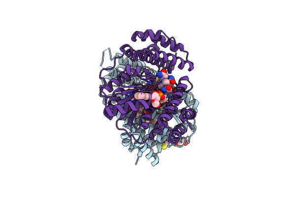
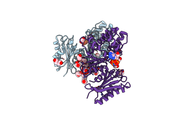
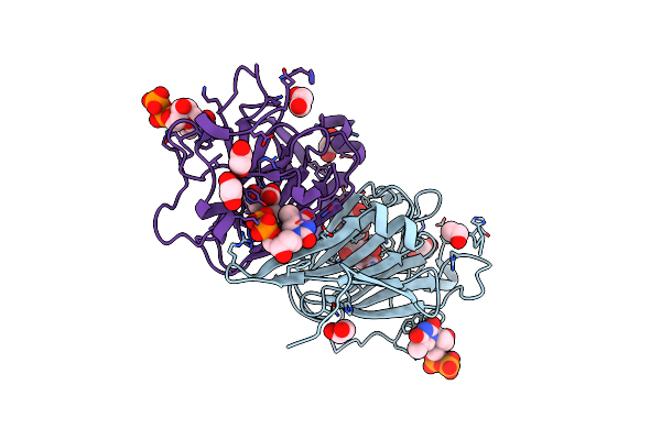
Closed Human Phosphoglycerate Kinase Complex With Bpg And Adp Produced By Cross-Soaking A Tsa Crystal
Organism: Homo sapiens
Method: X-RAY DIFFRACTION Resolution:1.76 Å Release Date: 2025-05-28 Classification: TRANSFERASE Ligands: ADP, X15, PEG, CL, MG, NA |
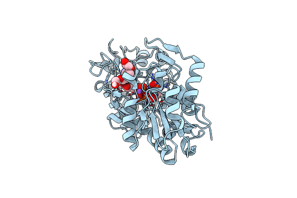 |
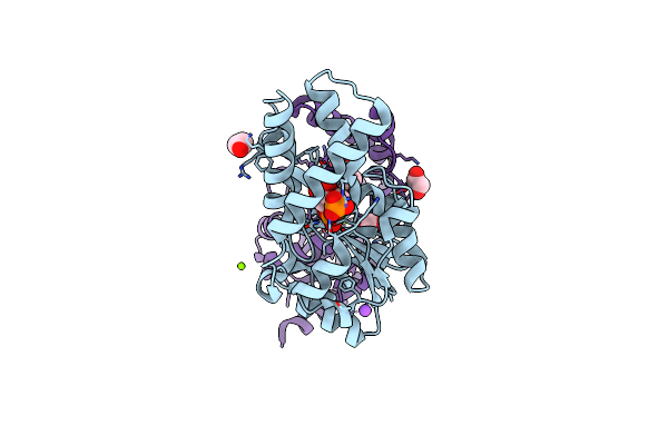
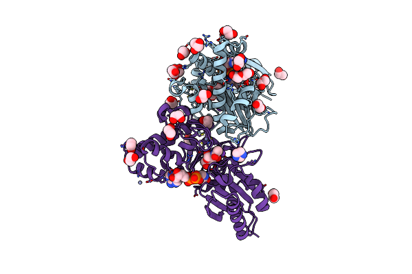
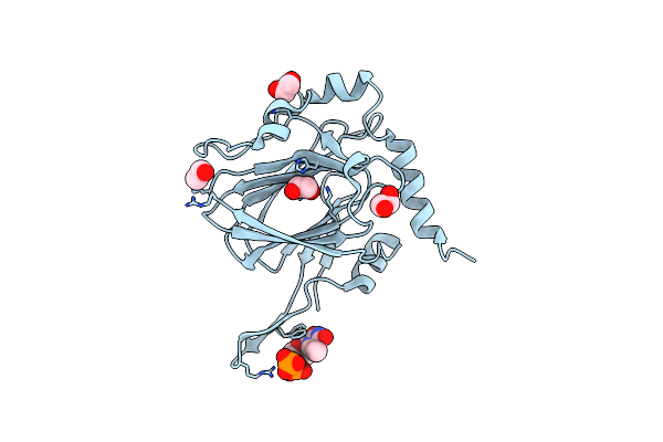
Human Phosphoglycerate Kinase In Complex With Atp And 3Pg Formed By Cross-Soaking A Tsa Crystal
Organism: Homo sapiens
Method: X-RAY DIFFRACTION Resolution:1.58 Å Release Date: 2025-05-28 Classification: TRANSFERASE |
 |
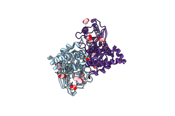
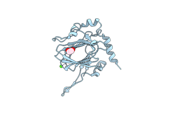
Substrate-Free D10N,P146A Variant Of Beta-Phosphoglucomutase From Lactococcus Lactis
Organism: Lactococcus lactis subsp. lactis il1403
Method: X-RAY DIFFRACTION Resolution:1.68 Å Release Date: 2024-08-07 Classification: ISOMERASE Ligands: MG, EDO, PO4, TRS |
 |
D10N Variant Of Beta-Phosphoglucomutase From Lactococcus Lactis In Complex With Fructose 1,6-Bisphosphate
Organism: Lactococcus lactis subsp. lactis il1403
Method: X-RAY DIFFRACTION Resolution:1.75 Å Release Date: 2024-08-07 Classification: ISOMERASE Ligands: FBP, MG, EDO |
 |
D10N,P146A Variant Of Beta-Phosphoglucomutase From Lactococcus Lactis In Complex With Fructose 1,6-Bisphosphate
Organism: Lactococcus lactis subsp. lactis il1403
Method: X-RAY DIFFRACTION Resolution:1.23 Å Release Date: 2024-08-07 Classification: ISOMERASE Ligands: FBP, MG, EDO |
 |
D10N,P146A Variant Of Beta-Phosphoglucomutase From Lactococcus Lactis In Complex With Native Beta-Glucose 1,6-Bisphosphate Intermediate
Organism: Lactococcus lactis subsp. lactis il1403
Method: X-RAY DIFFRACTION Resolution:1.01 Å Release Date: 2024-08-07 Classification: ISOMERASE Ligands: B16, MG, TRS, NA, EDO |
 |
Organism: Saccharothrix syringae
Method: X-RAY DIFFRACTION Resolution:1.70 Å Release Date: 2024-04-10 Classification: UNKNOWN FUNCTION Ligands: B12, 5AD, BLA, DIO, PEG |
 |
Organism: Saccharothrix syringae
Method: X-RAY DIFFRACTION Resolution:1.98 Å Release Date: 2024-04-10 Classification: UNKNOWN FUNCTION Ligands: B12, BLA, PEG |
 |
Organism: Acidimicrobiaceae bacterium
Method: X-RAY DIFFRACTION Resolution:2.30 Å Release Date: 2024-04-10 Classification: UNKNOWN FUNCTION Ligands: B12, 5AD, PEG, SCN |
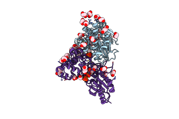 |
N-Acetylglucosamine Kinase From Plesiomonas Shigelloides Compexed With Alpha-N-Acetylglucosamine-6-Phosphate
Organism: Saccharomyces cerevisiae (strain atcc 204508 / s288c), Plesiomonas shigelloides 302-73
Method: X-RAY DIFFRACTION Resolution:1.75 Å Release Date: 2022-08-10 Classification: SUGAR BINDING PROTEIN Ligands: EDO, PEG, 4QY, ZN, K, PO4, CL, TRS |
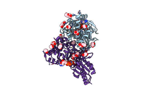 |
N-Acetylglucosamine Kinase From Plesiomonas Shigelloides Compexed With Alpha-N-Acetylglucosamine And Amp-Pnp Inhibitor
Organism: Saccharomyces cerevisiae (strain atcc 204508 / s288c), Plesiomonas shigelloides 302-73
Method: X-RAY DIFFRACTION Resolution:2.11 Å Release Date: 2022-08-10 Classification: SUGAR BINDING PROTEIN Ligands: ZN, ANP, PEG, PGE, NDG, EDO, K |
 |
N-Acetylglucosamine Kinase From Plesiomonas Shigelloides Compexed With Alpha-N-Acetylglucosamine
Organism: Saccharomyces cerevisiae (strain atcc 204508 / s288c), Plesiomonas shigelloides 302-73
Method: X-RAY DIFFRACTION Resolution:1.94 Å Release Date: 2022-08-10 Classification: SUGAR BINDING PROTEIN Ligands: MPD, EDO, PGE, NDG, ZN, K, CL, PEG |
 |
Structure Of N-Acetylglucosamine Kinase From Plesiomonas Shigelloides In Complex With Amp-Pnp In The Absence Of N-Acetylglucoseamine Substrate
Organism: Saccharomyces cerevisiae (strain atcc 204508 / s288c), Plesiomonas shigelloides 302-73
Method: X-RAY DIFFRACTION Resolution:2.20 Å Release Date: 2022-08-10 Classification: SUGAR BINDING PROTEIN Ligands: PEG, ANP, ZN, PGE, EDO |
 |
N-Acetylglucosamine Kinase From Plesiomonas Shigelloides Compexed With Alpha-N-Acetylglucosamine And Adp
Organism: Saccharomyces cerevisiae (strain atcc 204508 / s288c), Plesiomonas shigelloides 302-73
Method: X-RAY DIFFRACTION Resolution:1.57 Å Release Date: 2022-08-03 Classification: SUGAR BINDING PROTEIN Ligands: ADP, GOL, IMD, NDG, ZN, EDO, PEG, K, IPA |
 |
Native Structure Of N-Acetylglucosamine Kinase From Plesiomonas Shigelloides
Organism: Saccharomyces cerevisiae (strain atcc 204508 / s288c), Plesiomonas shigelloides 302-73
Method: X-RAY DIFFRACTION Resolution:1.70 Å Release Date: 2022-07-27 Classification: SUGAR BINDING PROTEIN Ligands: PEG, TRS, ZN, PGE, EDO, CL |
 |
Organism: Coxiella burnetii (strain rsa 493 / nine mile phase i)
Method: X-RAY DIFFRACTION Resolution:1.43 Å Release Date: 2022-04-20 Classification: SUGAR BINDING PROTEIN Ligands: XYP, XYS, NA, FLC |
 |
Organism: Coxiella burnetii (strain rsa 493 / nine mile phase i)
Method: X-RAY DIFFRACTION Resolution:1.87 Å Release Date: 2022-04-20 Classification: SUGAR BINDING PROTEIN Ligands: TYD, CIT, EDO |
 |
Organism: Streptomyces griseus
Method: X-RAY DIFFRACTION Resolution:1.90 Å Release Date: 2022-04-20 Classification: SUGAR BINDING PROTEIN Ligands: EDO, TYD, CL |
 |
Organism: Streptomyces griseus
Method: X-RAY DIFFRACTION Resolution:1.33 Å Release Date: 2022-04-20 Classification: SUGAR BINDING PROTEIN Ligands: EDO, CA |