
Search Count: 7
 |
Organism: Oryctolagus cuniculus
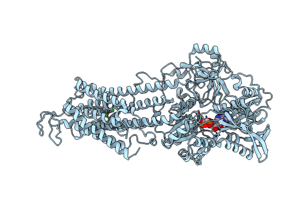
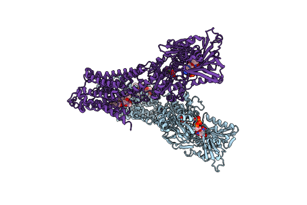
Method: X-RAY DIFFRACTION Resolution:3.20 Å Release Date: 2020-05-06 Classification: HYDROLASE Ligands: K, CA, ACP |
 |
Structure Of The (Sr) Ca2+-Atpase Bound To A Tetrahydrocarbazole And Tnp-Atp
Organism: Oryctolagus cuniculus
Method: X-RAY DIFFRACTION Resolution:3.00 Å Release Date: 2018-01-10 Classification: HYDROLASE Ligands: 128, K, PCW, 8T8 |
 |
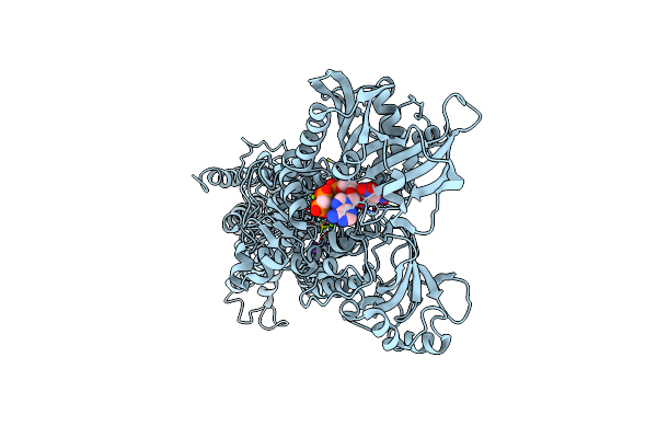
Crystal Structure Of The (Sr) Calcium Atpase E2-Vanadate Complex Bound To Thapsigargin And Tnp-Amppcp
Organism: Oryctolagus cuniculus
Method: X-RAY DIFFRACTION Resolution:3.05 Å Release Date: 2016-04-13 Classification: HYDROLASE Ligands: TG1, VN4, DL5, MG, K, CL |
 |
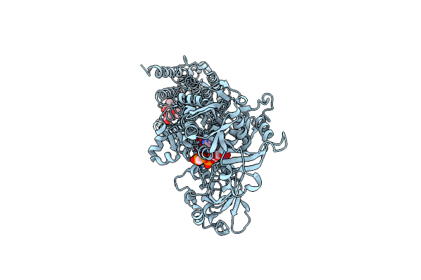
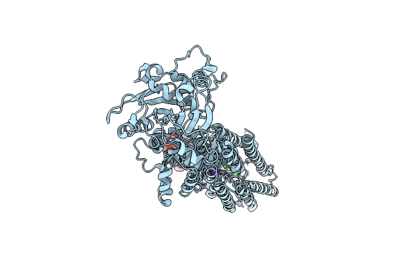
Crystal Structure Of The (Sr) Calcium Atpase E2.Bef3- Complex Bound To Tnp-Amppcp
Organism: Oryctolagus cuniculus
Method: X-RAY DIFFRACTION Resolution:3.05 Å Release Date: 2016-04-13 Classification: HYDROLASE Ligands: DL5, K, MG |
 |
Crystal Structure Of The (Sr) Calcium Atpase E2-Vanadate Complex Bound To Thapsigargin And Tnp-Atp
Organism: Oryctolagus cuniculus
Method: X-RAY DIFFRACTION Resolution:3.30 Å Release Date: 2016-04-13 Classification: HYDROLASE Ligands: TG1, VN4, 128, MG, K, CL |
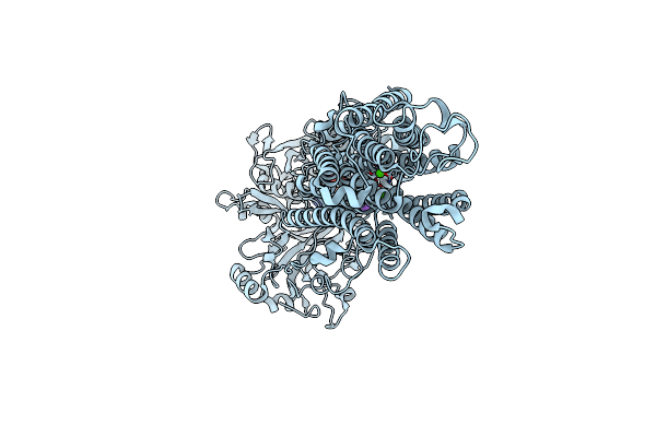 |
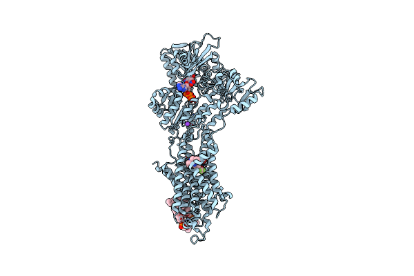
Crystal Structure Of The Sr Ca2+-Atpase In The Ca2-E1-Mgamppcp Form Determined By Serial Femtosecond Crystallography Using An X-Ray Free-Electron Laser.
Organism: Oryctolagus cuniculus
Method: X-RAY DIFFRACTION Resolution:2.80 Å Release Date: 2015-06-10 Classification: HYDROLASE Ligands: CA, K, ACP |
 |
Organism: Oryctolagus cuniculus
Method: X-RAY DIFFRACTION Resolution:3.50 Å Release Date: 2013-12-18 Classification: HYDROLASE Ligands: CA, PCW, K |