
Search Count: 37
 |
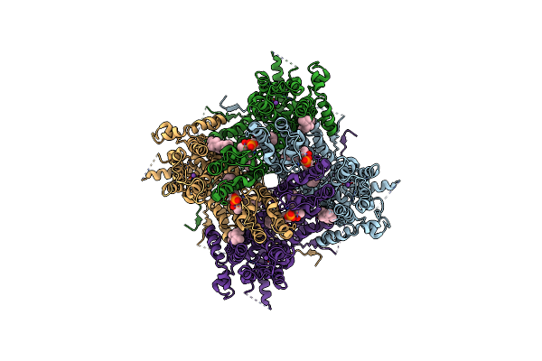
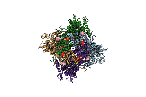
Structure Of Sars-Cov-2 Orf3A In Late Endosome/Lysosome-Like Membrane Environment, Msp1D1 Nanodisc
Organism: Severe acute respiratory syndrome coronavirus 2
Method: ELECTRON MICROSCOPY Release Date: 2023-02-08 Classification: VIRAL PROTEIN Ligands: PEE |
 |
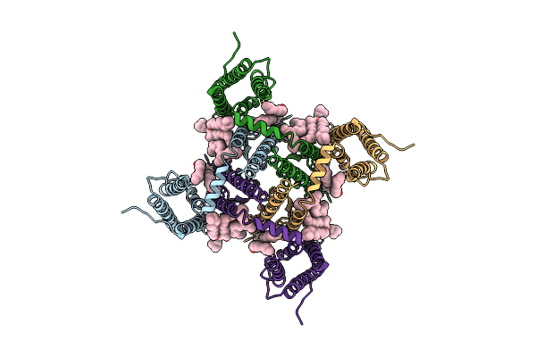
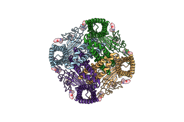
Structure Of Sars-Cov-1 Orf3A In Late Endosome/Lysosome-Like Environment, Msp1D1 Nanodisc
Organism: Severe acute respiratory syndrome coronavirus, Homo sapiens
Method: ELECTRON MICROSCOPY Release Date: 2023-02-08 Classification: VIRAL PROTEIN Ligands: PEE |
 |
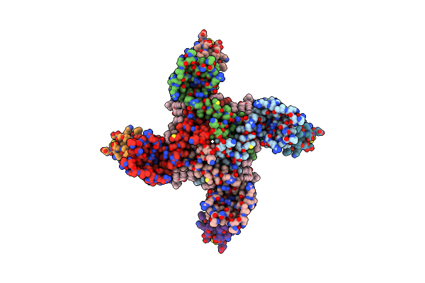
Structure Of Sars-Cov-2 Orf3A In Plasma Membrane-Like Environment, Msp1D1 Nanodisc
Organism: Severe acute respiratory syndrome coronavirus 2
Method: ELECTRON MICROSCOPY Release Date: 2023-02-08 Classification: VIRAL PROTEIN Ligands: PEE |
 |
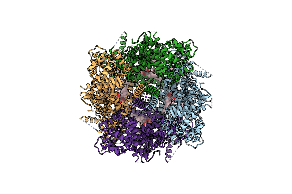
Structure Of Sars-Cov-2 Orf3A In Late Endosome/Lysosome-Like Environment, Saposin A Nanodisc
Organism: Severe acute respiratory syndrome coronavirus 2, Homo sapiens
Method: ELECTRON MICROSCOPY Release Date: 2023-02-08 Classification: VIRAL PROTEIN Ligands: PEE |
 |
Organism: Mus musculus
Method: ELECTRON MICROSCOPY Release Date: 2020-10-21 Classification: MEMBRANE PROTEIN Ligands: Y01, LPP, NA |
 |
Organism: Bacillus halodurans c-125
Method: ELECTRON MICROSCOPY Release Date: 2020-06-24 Classification: MEMBRANE PROTEIN Ligands: POV, NA |
 |
Organism: Bacillus halodurans c-125
Method: ELECTRON MICROSCOPY Release Date: 2020-06-24 Classification: MEMBRANE PROTEIN Ligands: POV |
 |
Organism: Bacillus halodurans c-125, Homo sapiens
Method: ELECTRON MICROSCOPY Release Date: 2020-06-24 Classification: MEMBRANE PROTEIN Ligands: POV |
 |
Organism: Bacillus halodurans (strain atcc baa-125 / dsm 18197 / ferm 7344 / jcm 9153 / c-125), Homo sapiens, Cyriopagopus schmidti
Method: ELECTRON MICROSCOPY Release Date: 2020-06-24 Classification: TRANSPORT PROTEIN/TOXIN Ligands: POV |
 |
Organism: Mus musculus
Method: ELECTRON MICROSCOPY Release Date: 2019-08-07 Classification: MEMBRANE PROTEIN |
 |
Organism: Homo sapiens
Method: ELECTRON MICROSCOPY Release Date: 2018-12-19 Classification: MEMBRANE PROTEIN |
 |
Organism: Mus musculus
Method: ELECTRON MICROSCOPY Release Date: 2018-10-17 Classification: TRANSFERASE Ligands: Y01 |
 |
Organism: Mus musculus
Method: ELECTRON MICROSCOPY Release Date: 2018-08-15 Classification: MEMBRANE PROTEIN Ligands: MG, Y01 |
 |
Organism: Mus musculus
Method: ELECTRON MICROSCOPY Release Date: 2018-08-15 Classification: MEMBRANE PROTEIN |
 |
Organism: Homo sapiens
Method: ELECTRON MICROSCOPY Release Date: 2018-07-25 Classification: MEMBRANE PROTEIN Ligands: NAG |
 |
Organism: Mus musculus
Method: ELECTRON MICROSCOPY Release Date: 2018-04-18 Classification: MEMBRANE PROTEIN Ligands: Y01, LPP, NA |
 |
Pseudomonas Fluorescens Kynurenine 3-Monooxygenase (Kmo) In Complex With 3-(5-Chloro-6-Ethoxy-2-Oxo-2,3-Dihydro-1,3-Benzoxazol-3-Yl)Propanoic Acid
Organism: Pseudomonas fluorescens
Method: X-RAY DIFFRACTION Resolution:1.82 Å Release Date: 2017-04-19 Classification: OXIDOREDUCTASE Ligands: FAD, CL, 8EQ, GOL |
 |
Pseudomonas Fluorescens Kynurenine 3-Monooxygenase (Kmo) In Complex With 3-(5-Chloro-6-Cyclopropoxy-2-Oxo-2,3-Dihydro-1,3-Benzoxazol-3-Yl)Propanoic Acid
Organism: Pseudomonas fluorescens
Method: X-RAY DIFFRACTION Resolution:1.71 Å Release Date: 2017-04-19 Classification: OXIDOREDUCTASE Ligands: FAD, CL, FYK, GOL |
 |
Pseudomonas Fluorescens Kynurenine 3-Monooxygenase (Kmo) In Complex With 3-[5-Chloro-6-(Cyclobutylmethoxy)-2-Oxo-2,3-Dihydro-1,3-Benzoxazol-3-Yl]Propanoic Acid
Organism: Pseudomonas fluorescens
Method: X-RAY DIFFRACTION Resolution:1.82 Å Release Date: 2017-04-19 Classification: OXIDOREDUCTASE Ligands: FAD, CL, OK1, GOL |
 |
Cryo-Em Structure Of Polycystic Kidney Disease Protein 2 (Pkd2), Residues 198-703
Organism: Homo sapiens
Method: ELECTRON MICROSCOPY Release Date: 2016-11-02 Classification: METAL TRANSPORT Ligands: NAG |