
Search Count: 13
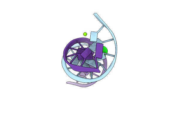 |
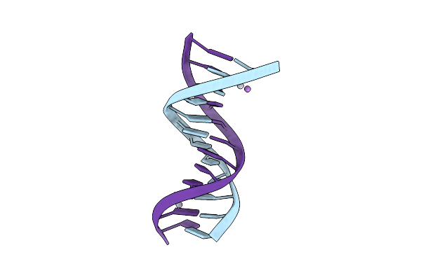
Crystal Structure Of Transplatin/B-Dna Adduct Obtained Upon 7 Days Of Soaking
Organism: Synthetic construct
Method: X-RAY DIFFRACTION Resolution:1.40 Å Release Date: 2024-02-28 Classification: DNA Ligands: MG, PT, NH3, CL |
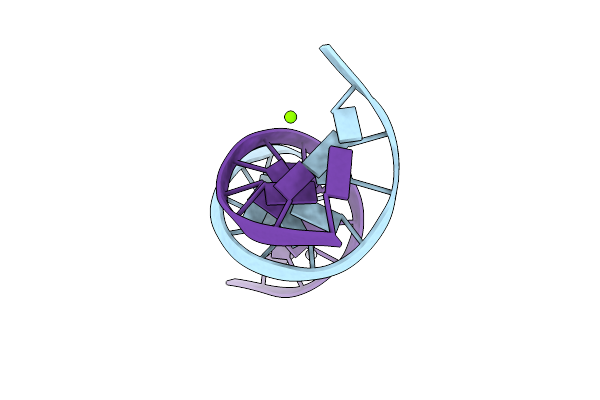 |
Crystal Structure Of Transplatin/B-Dna Adduct Obtained Upon 48 H Of Soaking
Organism: Dna molecule
Method: X-RAY DIFFRACTION Resolution:1.42 Å Release Date: 2024-02-07 Classification: DNA Ligands: MG, NH3, PT |
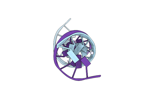 |
Organism: Synthetic construct
Method: X-RAY DIFFRACTION Resolution:2.31 Å Release Date: 2024-01-31 Classification: DNA Ligands: PT |
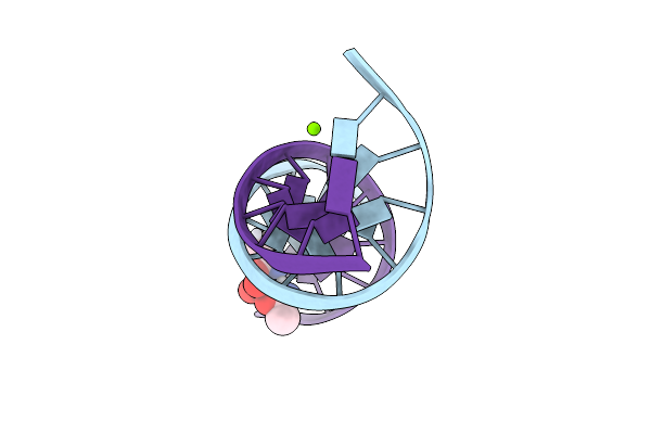 |
Crystal Structure Of Arsenoplatin-1/B-Dna Adduct Obtained Upon 4 H Of Soaking
Organism: Synthetic construct
Method: X-RAY DIFFRACTION Resolution:1.52 Å Release Date: 2024-01-31 Classification: DNA Ligands: PT, MG, A6R |
 |
Crystal Structure Of Arsenoplatin-1/B-Dna Adduct Obtained Upon 48 H Of Soaking
Organism: Synthetic construct
Method: X-RAY DIFFRACTION Resolution:2.51 Å Release Date: 2024-01-31 Classification: DNA Ligands: PT, A6R |
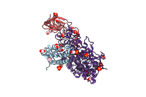 |
X-Ray Crystal Structure Of Sporosarcina Pasteurii Urease Inhibited By Ag(Pet3)Cl Determined At 1.72 Angstroms
Organism: Sporosarcina pasteurii
Method: X-RAY DIFFRACTION Resolution:1.72 Å Release Date: 2021-10-13 Classification: HYDROLASE Ligands: EDO, SO4, NI, O, AG |
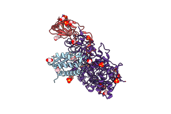 |
X-Ray Crystal Structure Of Sporosarcina Pasteurii Urease Inhibited By Ag(Pet3)Br Determined At 1.63 Angstroms
Organism: Sporosarcina pasteurii
Method: X-RAY DIFFRACTION Resolution:1.63 Å Release Date: 2021-10-13 Classification: HYDROLASE Ligands: EDO, SO4, NI, O, AG |
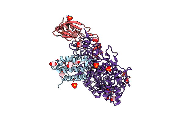 |
X-Ray Crystal Structure Of Sporosarcina Pasteurii Urease Inhibited By Ag(Pet3)2No3 Determined At 1.97 Angstroms
Organism: Sporosarcina pasteurii
Method: X-RAY DIFFRACTION Resolution:1.97 Å Release Date: 2021-10-13 Classification: HYDROLASE Ligands: EDO, SO4, NI, O, AG |
 |
Organism: Equus caballus
Method: X-RAY DIFFRACTION Resolution:1.50 Å Release Date: 2021-03-17 Classification: TRANSPORT PROTEIN |
 |
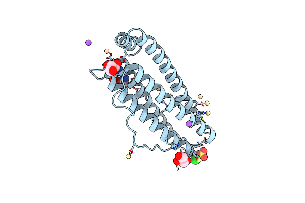
X-Ray Structure Of The Adduct Formed Upon Treating Lysozyme With An Aged Solution Of Arsenoplatin-1
Organism: Gallus gallus
Method: X-RAY DIFFRACTION Resolution:2.15 Å Release Date: 2020-12-23 Classification: HYDROLASE Ligands: NO3, EDO, A6R, DMS, NA |
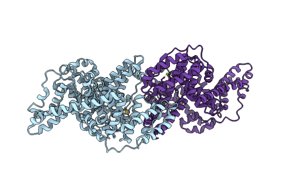 |
The X-Ray Structure Of The Gold/Serum Albumin Adduct Obtained Upon Reaction Of The Protein With Aul12, A Gold(Iii) Dithiocarbamate Complex
Organism: Bos taurus
Method: X-RAY DIFFRACTION Resolution:3.21 Å Release Date: 2019-08-14 Classification: TRANSPORT PROTEIN Ligands: MG, AU |
 |
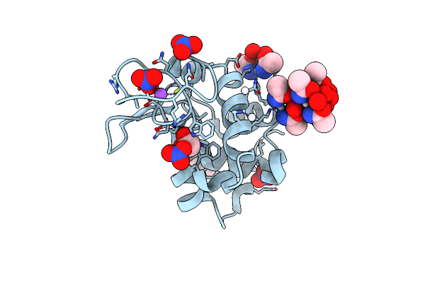
X-Ray Structure Of The Adduct Formed Upon Reaction Of Ribonuclease A With A Tetranuclear Pt-Thiosemicarbazone Compound
Organism: Bos taurus
Method: X-RAY DIFFRACTION Resolution:1.78 Å Release Date: 2018-08-29 Classification: HYDROLASE Ligands: PT, DMS |
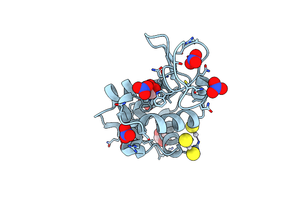 |
X-Ray Structure Of The Adduct Formed Upon Reaction Of Hen Egg White Lysozyme With A Tetranuclear Pt-Thiosemicarbazone Compound
Organism: Gallus gallus
Method: X-RAY DIFFRACTION Resolution:1.78 Å Release Date: 2018-08-29 Classification: HYDROLASE Ligands: EDO, NO3, DMS, PT |