
Search Count: 104
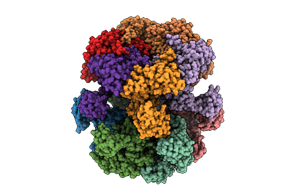 |
Organism: Homo sapiens
Method: ELECTRON MICROSCOPY Resolution:3.50 Å Release Date: 2025-12-24 Classification: CHAPERONE Ligands: ADP |
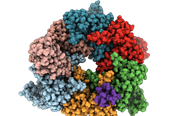 |
Organism: Homo sapiens
Method: ELECTRON MICROSCOPY Release Date: 2025-11-19 Classification: CHAPERONE Ligands: ADP |
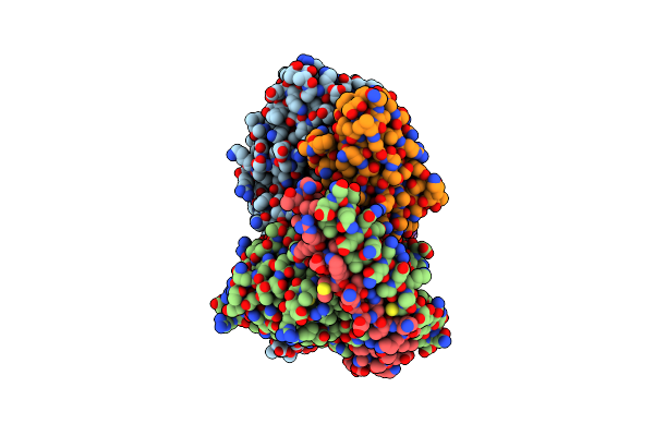 |
Organism: Homo sapiens, Lama glama, Escherichia coli
Method: ELECTRON MICROSCOPY Release Date: 2024-10-09 Classification: MEMBRANE PROTEIN Ligands: FWX, CLR |
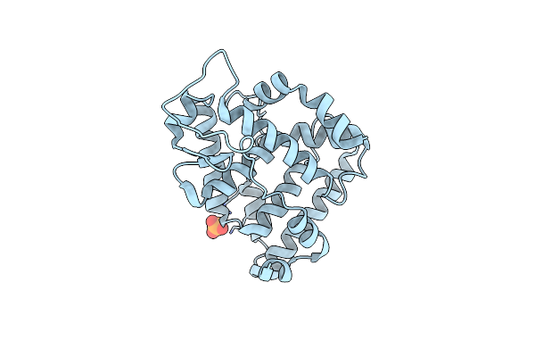 |
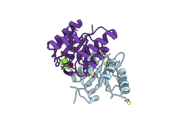
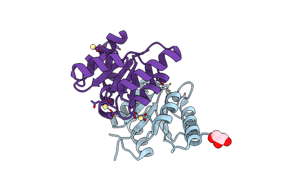
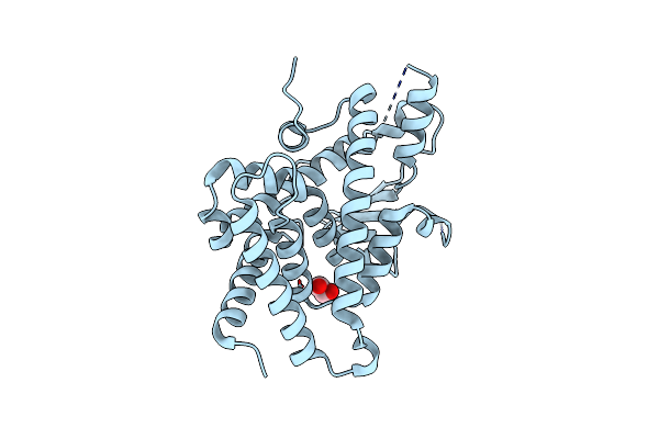
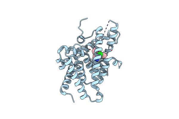
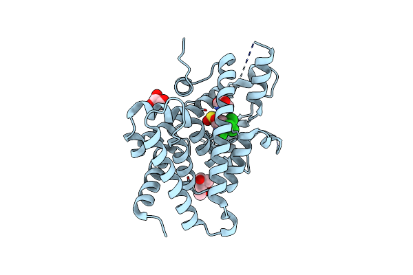
The Crystallographic Structure Of The Ligand Binding Domain Of The Nr7 Nuclear Receptor From The Amphioxus Branchiostoma Lanceolatum
Organism: Branchiostoma lanceolatum
Method: X-RAY DIFFRACTION Resolution:2.00 Å Release Date: 2022-09-14 Classification: TRANSCRIPTION Ligands: CL, PO4 |
 |
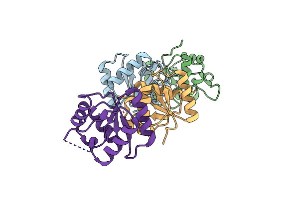
Heterodimeric Murine Trna-Guanine Transglycosylase In Complex With Queuine And In The Presence Of Anderson-Evans Type (Tew) And Strandberg Type Polyoxometalate (Pom)
Organism: Mus musculus
Method: X-RAY DIFFRACTION Resolution:2.60 Å Release Date: 2022-07-13 Classification: TRANSFERASE Ligands: TEW, ZN, QEI, 2I2 |
 |
Organism: Pseudomonas aeruginosa pao1
Method: X-RAY DIFFRACTION Resolution:2.64 Å Release Date: 2022-07-13 Classification: SIGNALING PROTEIN Ligands: CA, PGR |
 |
Organism: Pseudomonas aeruginosa
Method: X-RAY DIFFRACTION Resolution:1.87 Å Release Date: 2022-07-06 Classification: SIGNALING PROTEIN Ligands: CD, BEF, MG |
 |
Organism: Pseudomonas aeruginosa
Method: X-RAY DIFFRACTION Resolution:1.45 Å Release Date: 2022-07-06 Classification: SIGNALING PROTEIN Ligands: CD, GOL |
 |
Organism: Mus musculus
Method: X-RAY DIFFRACTION Resolution:1.90 Å Release Date: 2022-06-22 Classification: TRANSFERASE Ligands: SO4, ZN, PG4, PGE, P6G |
 |
Organism: Mus musculus
Method: X-RAY DIFFRACTION Resolution:2.10 Å Release Date: 2022-06-22 Classification: TRANSFERASE Ligands: ZN, SO4, DMS, QEI |
 |
Heterodimeric Murine Trna-Guanine Transglycosylase In The Presence Of Anderson-Evans Type (Tew) And Strandberg Type Polyoxometalate (Pom)
Organism: Mus musculus
Method: X-RAY DIFFRACTION Resolution:2.60 Å Release Date: 2022-06-22 Classification: TRANSFERASE Ligands: ZN, TEW, 2I2 |
 |
Organism: Mus musculus
Method: X-RAY DIFFRACTION Resolution:1.65 Å Release Date: 2021-12-08 Classification: TRANSFERASE Ligands: ZN, SO4, PEG, PG4, PGE, P6G, CIT |
 |
Organism: Geobacillus stearothermophilus
Method: X-RAY DIFFRACTION Resolution:2.14 Å Release Date: 2021-03-17 Classification: HYDROLASE Ligands: MG |
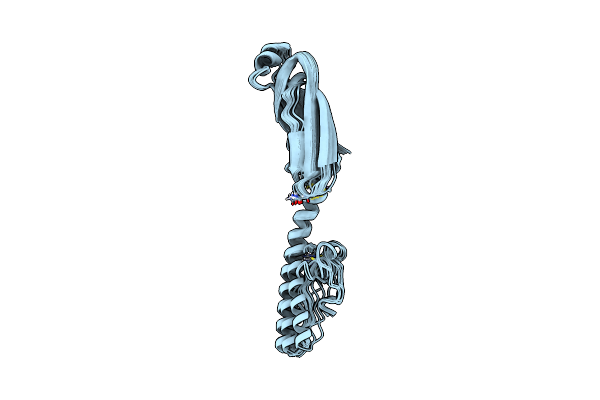 |
Full Structure Of Rymv P1 Protein, Derived From Crystallographic And Nmr Data.
Organism: Rice yellow mottle virus
Method: SOLUTION NMR Release Date: 2021-02-03 Classification: VIRAL PROTEIN Ligands: ZN |
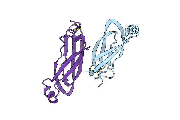 |
Nt Part Crystal Structure Of The Rymv-Encoded Viral Rna Silencing Suppressor P1
Organism: Rice yellow mottle virus
Method: X-RAY DIFFRACTION Resolution:2.10 Å Release Date: 2021-01-27 Classification: RNA BINDING PROTEIN Ligands: ZN |
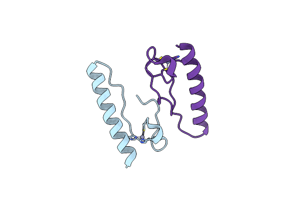 |
Ct Part Crystal Structure Of The Rymv-Encoded Viral Rna Silencing Suppressor P1
Organism: Rice yellow mottle virus
Method: X-RAY DIFFRACTION Resolution:1.98 Å Release Date: 2021-01-27 Classification: RNA BINDING PROTEIN Ligands: ZN |
 |
Organism: Homo sapiens
Method: X-RAY DIFFRACTION Resolution:2.15 Å Release Date: 2021-01-13 Classification: NUCLEAR PROTEIN Ligands: EDO |
 |
Organism: Homo sapiens
Method: X-RAY DIFFRACTION Resolution:2.25 Å Release Date: 2021-01-13 Classification: NUCLEAR PROTEIN Ligands: S6H, IPA, GOL |
 |
Organism: Homo sapiens
Method: X-RAY DIFFRACTION Resolution:2.26 Å Release Date: 2021-01-13 Classification: NUCLEAR PROTEIN Ligands: CL6 |
 |
Organism: Homo sapiens
Method: X-RAY DIFFRACTION Resolution:2.55 Å Release Date: 2021-01-13 Classification: NUCLEAR PROTEIN Ligands: MPD, S68, GOL |