
Search Count: 24
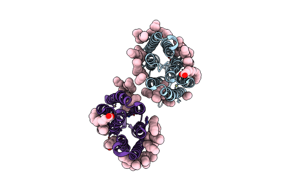 |
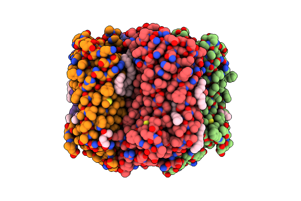
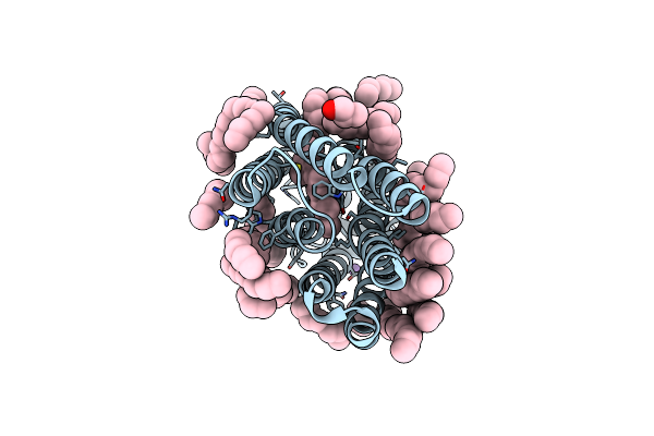
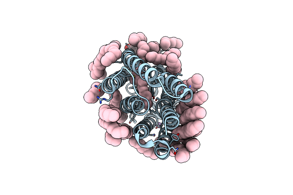
Crystal Structure Of A Light-Driven Proton Pump Lr (Mac) From Leptosphaeria Maculans
Organism: Leptosphaeria maculans
Method: X-RAY DIFFRACTION Resolution:2.20 Å Release Date: 2021-07-07 Classification: MEMBRANE PROTEIN Ligands: LFA, OLA |
 |
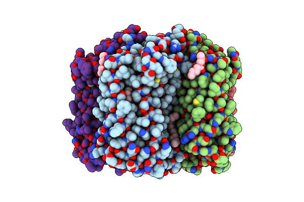
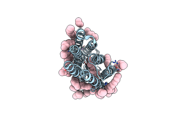
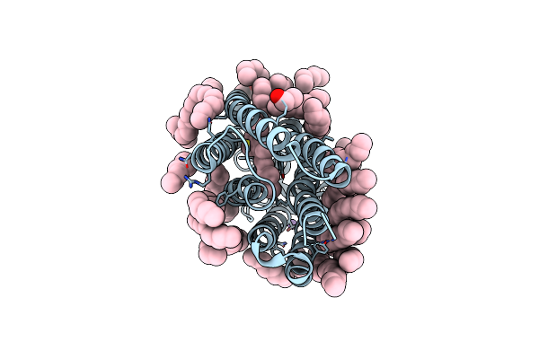
Crystal Structure Of The N112A Mutant Of The Light-Driven Sodium Pump Kr2 In The Pentameric Form, Ph 8.0
Organism: Dokdonia eikasta
Method: X-RAY DIFFRACTION Resolution:2.40 Å Release Date: 2020-06-17 Classification: MEMBRANE PROTEIN Ligands: OLC, OLA, LFA, BOG, RET, NA |
 |
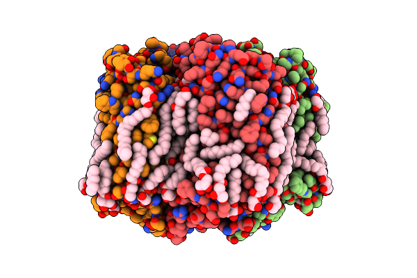
Crystal Structure Of The C14 Ring Of The F1Fo Atp Synthase From Spinach Chloroplast
Organism: Spinacia oleracea
Method: X-RAY DIFFRACTION Resolution:2.30 Å Release Date: 2019-12-25 Classification: MEMBRANE PROTEIN Ligands: LFA, OLC |
 |
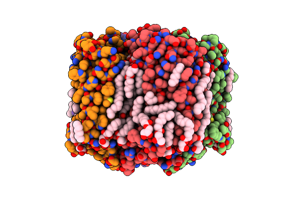
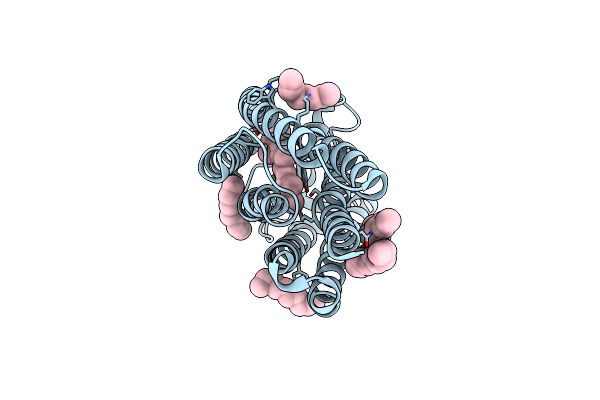
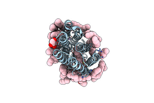
Crystal Structure Of The Light-Driven Sodium Pump Kr2 In The Pentameric Form, Ph 6.0
Organism: Dokdonia eikasta
Method: X-RAY DIFFRACTION Resolution:2.70 Å Release Date: 2019-04-24 Classification: MEMBRANE PROTEIN Ligands: OLC, LFA, NA, BOG, RET |
 |
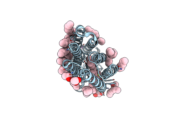
Crystal Structure Of The Light-Driven Sodium Pump Kr2 In The Pentameric Form, Ph 5.0
Organism: Dokdonia eikasta
Method: X-RAY DIFFRACTION Resolution:2.60 Å Release Date: 2019-04-24 Classification: MEMBRANE PROTEIN Ligands: LFA, OLC, NA, RET |
 |
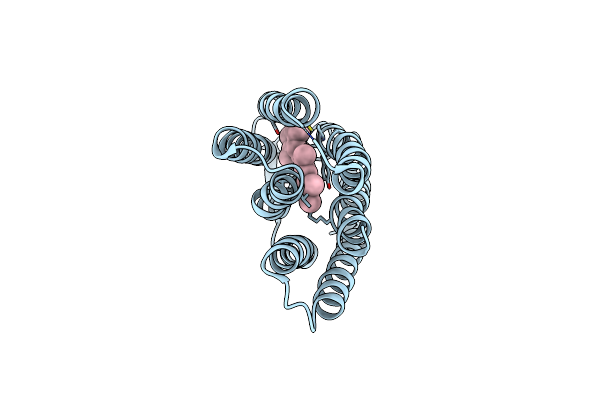
Crystal Structure Of The Light-Driven Sodium Pump Kr2 In The Pentameric "Dry" Form
Organism: Dokdonia eikasta
Method: X-RAY DIFFRACTION Resolution:3.00 Å Release Date: 2019-04-24 Classification: MEMBRANE PROTEIN |
 |
Crystal Structure Of The Light-Driven Sodium Pump Kr2 In The Pentameric "Wet" Form
Organism: Dokdonia eikasta
Method: X-RAY DIFFRACTION Resolution:2.80 Å Release Date: 2019-04-24 Classification: MEMBRANE PROTEIN Ligands: OLC, LFA, NA, BOG, RET |
 |
Crystal Structure Of The Potassium-Pumping G263F Mutant Of The Light-Driven Sodium Pump Kr2 In The Pentameric Form, Ph 8.0
Organism: Dokdonia eikasta
Method: X-RAY DIFFRACTION Resolution:2.40 Å Release Date: 2019-04-24 Classification: MEMBRANE PROTEIN Ligands: OLC, LFA, NA, BOG |
 |
Crystal Structure Of The Potassium-Pumping S254A Mutant Of The Light-Driven Sodium Pump Kr2 In The Pentameric Form, Ph 8.0
Organism: Dokdonia eikasta
Method: X-RAY DIFFRACTION Resolution:2.40 Å Release Date: 2019-04-24 Classification: MEMBRANE PROTEIN Ligands: OLC, LFA, NA, BOG, RET |
 |
Crystal Structure Of The Light-Driven Sodium Pump Kr2 In The Monomeric Form, Ph 6.0
Organism: Dokdonia eikasta
Method: X-RAY DIFFRACTION Resolution:2.30 Å Release Date: 2019-04-24 Classification: MEMBRANE PROTEIN |
 |
Crystal Structure Of The Light-Driven Sodium Pump Kr2 In The Monomeric Form, Ph 8.0
Organism: Dokdonia eikasta
Method: X-RAY DIFFRACTION Resolution:1.80 Å Release Date: 2019-04-24 Classification: MEMBRANE PROTEIN Ligands: LFA, NA, RET |
 |
Crystal Structure Of The Light-Driven Sodium Pump Kr2 In The Monomeric Form, Ph 8.9
Organism: Dokdonia eikasta
Method: X-RAY DIFFRACTION Resolution:2.60 Å Release Date: 2019-04-24 Classification: MEMBRANE PROTEIN Ligands: LFA, RET |
 |
Crystal Structure Of The Y154F Mutant Of The Light-Driven Sodium Pump Kr2 In The Monomeric Form, Ph 8.0
Organism: Dokdonia eikasta
Method: X-RAY DIFFRACTION Resolution:1.80 Å Release Date: 2019-04-24 Classification: MEMBRANE PROTEIN Ligands: LFA, GOL, NA, RET |
 |
Crystal Structure Of The H30K Mutant Of The Light-Driven Sodium Pump Kr2 In The Monomeric Form, Ph 8.0
Organism: Dokdonia eikasta
Method: X-RAY DIFFRACTION Resolution:2.20 Å Release Date: 2019-04-24 Classification: MEMBRANE PROTEIN |
 |
Crystal Structure Of The Potassium-Pumping S254A Mutant Of The Light-Driven Sodium Pump Kr2 In The Monomeric Form, Ph 4.3
Organism: Dokdonia eikasta
Method: X-RAY DIFFRACTION Resolution:2.10 Å Release Date: 2019-04-24 Classification: MEMBRANE PROTEIN |
 |
Crystal Structure Of The Potassium-Pumping G263F Mutant Of The Light-Driven Sodium Pump Kr2 In The Monomeric Form, Ph 4.3
Organism: Dokdonia eikasta
Method: X-RAY DIFFRACTION Resolution:2.00 Å Release Date: 2019-04-24 Classification: MEMBRANE PROTEIN |
 |
Structure Of Bacteriorhdopsin Transferred From Amphipol A8-35 To A Lipidic Mesophase
Organism: Halobacterium sp.
Method: X-RAY DIFFRACTION Resolution:2.00 Å Release Date: 2014-10-01 Classification: TRANSPORT PROTEIN Ligands: RET |
 |
Organism: Exiguobacterium sibiricum
Method: X-RAY DIFFRACTION Resolution:2.30 Å Release Date: 2013-07-17 Classification: PROTON TRANSPORT Ligands: RET, LFA |
 |
Refined Structure Of Glycophorin A Transmembrane Segment Dimer In Dpc Micelles
Organism: Homo sapiens
Method: SOLUTION NMR Release Date: 2010-09-22 Classification: MEMBRANE PROTEIN |
 |
Spatial Structure Of The Dimeric Transmembrane Domain Of Glycophorin A In Bicelles Soluton
Organism: Homo sapiens
Method: SOLUTION NMR Release Date: 2010-09-22 Classification: MEMBRANE PROTEIN |