
Search Count: 117
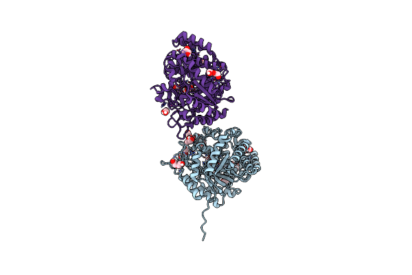 |
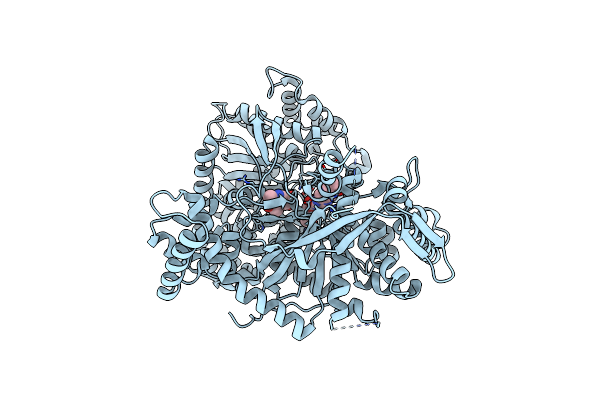
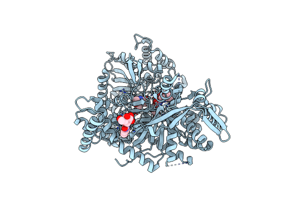
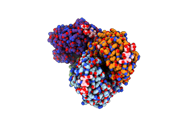
The Crystal Structure Of Beta-Glucosidase From The Thermophilic Bacterium Caldicellulosiruptor Saccharolyticus Determined At 1.47 A Resolution
Organism: Caldicellulosiruptor saccharolyticus dsm 8903
Method: X-RAY DIFFRACTION Resolution:1.47 Å Release Date: 2024-10-16 Classification: HYDROLASE Ligands: GOL, EDO, PGE, 1PE, PEG, CL |
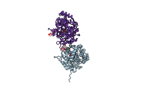 |
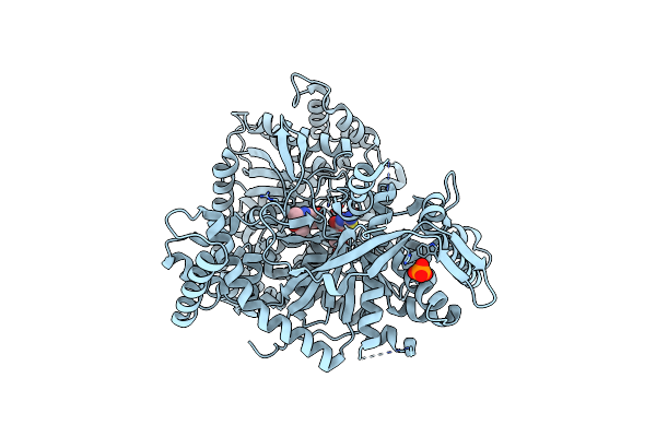
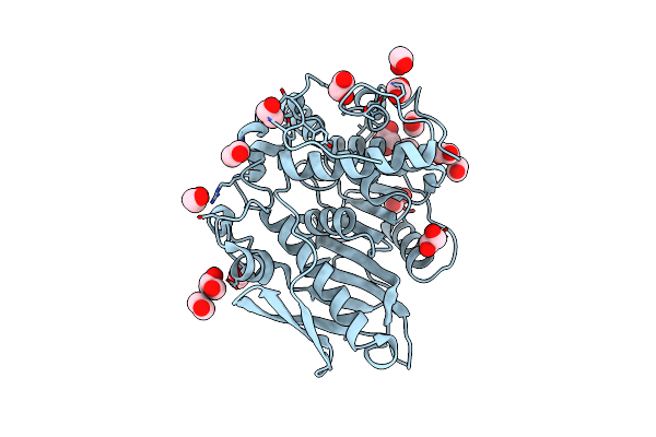
The Crystal Structure Of Beta-Glucosidase From The Thermophilic Bacterium Caldicellulosiruptor Saccharolyticus In Complex With Beta-D-Glucose Determined At 1.95 A Resolution
Organism: Caldicellulosiruptor saccharolyticus dsm 8903
Method: X-RAY DIFFRACTION Resolution:1.95 Å Release Date: 2024-10-16 Classification: HYDROLASE Ligands: BGC, 1PE, PEG, CL |
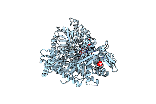 |
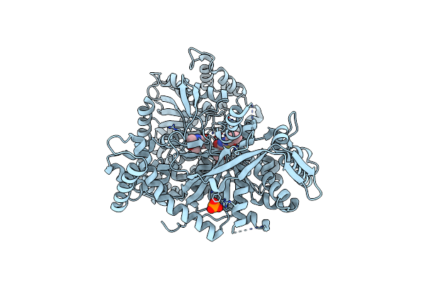
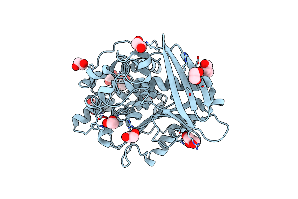
A Glucose-Based Molecular Rotor Probes The Catalytic Site Of Glycogen Phosphorylase.
Organism: Oryctolagus cuniculus
Method: X-RAY DIFFRACTION Resolution:1.80 Å Release Date: 2022-03-02 Classification: SUGAR BINDING PROTEIN Ligands: I0F, CO3, BME |
 |
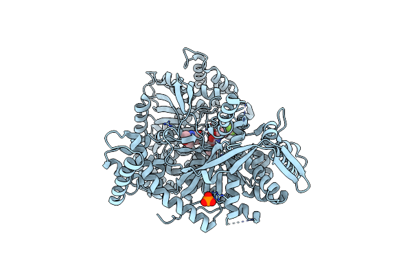
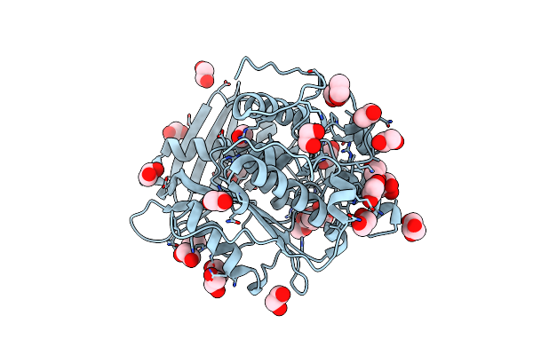
Organism: Fusarium oxysporum
Method: X-RAY DIFFRACTION Resolution:2.30 Å Release Date: 2019-01-30 Classification: HYDROLASE Ligands: NAG, CA |
 |
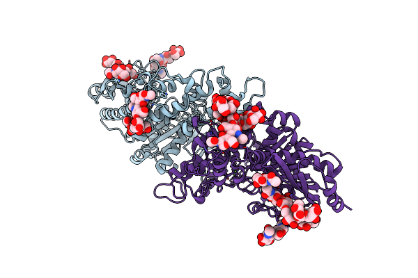
A Potent Fluorescent Inhibitor Of Glycogen Phosphorylase As A Catalytic Site Probe.
Organism: Oryctolagus cuniculus
Method: X-RAY DIFFRACTION Resolution:1.78 Å Release Date: 2017-11-29 Classification: FLUORESCENT PROTEIN Ligands: PLP, 7LS |
 |
Glucopyranosylidene-Spiro-Iminothiazolidinone, A New Bicyclic Ring System: Synthesis, Derivatization, And Evaluation As Glycogen Phosphorylase Inhibitors By Enzyme Kinetic And Crystallographic Methods
Organism: Oryctolagus cuniculus
Method: X-RAY DIFFRACTION Resolution:1.95 Å Release Date: 2014-08-06 Classification: TRANSFERASE Ligands: MIF, PLP, PO4 |
 |
Glucopyranosylidene-Spiro-Iminothiazolidinone, A New Bicyclic Ring System: Synthesis, Derivatization, And Evaluation As Glycogen Phosphorylase Inhibitors By Enzyme Kinetic And Crystallographic Methods
Organism: Oryctolagus cuniculus
Method: X-RAY DIFFRACTION Resolution:2.10 Å Release Date: 2014-08-06 Classification: TRANSFERASE Ligands: DTT, DMS, M8P, PLP |
 |
Glucopyranosylidene-Spiro-Iminothiazolidinone, A New Bicyclic Ring System: Synthesis, Derivatization, And Evaluation As Glycogen Phosphorylase Inhibitors By Enzyme Kinetic And Crystallographic Methods
Organism: Oryctolagus cuniculus
Method: X-RAY DIFFRACTION Resolution:1.90 Å Release Date: 2014-08-06 Classification: TRANSFERASE Ligands: M7C, PLP, PO4 |
 |
Rabbit Muscle Glycogen Phosphorylase B In Complex With N- Cyclohexancarbonyl-N-Beta-D-Glucopyranosyl Urea Determined At 1.83 A Resolution
Organism: Oryctolagus cuniculus
Method: X-RAY DIFFRACTION Resolution:1.83 Å Release Date: 2013-12-11 Classification: TRANSFERASE Ligands: PLP, F58 |
 |
Rabbit Muscle Glycogen Phosphorylase B In Complex With N-(4- Trifluoromethyl-Benzoyl)-N-Beta-D-Glucopyranosyl Urea Determined At 2. 15 A Resolution
Organism: Oryctolagus cuniculus
Method: X-RAY DIFFRACTION Resolution:2.15 Å Release Date: 2013-12-11 Classification: TRANSFERASE Ligands: PLP, 62N, PO4 |
 |
Rabbit Muscle Glycogen Phosphorylase B In Complex With N-(4-Tert- Butyl-Benzoyl)-N-Beta-D-Glucopyranosyl Urea Determined At 2.07 A Resolution
Organism: Oryctolagus cuniculus
Method: X-RAY DIFFRACTION Resolution:2.07 Å Release Date: 2013-12-11 Classification: TRANSFERASE Ligands: IMP, PLP, F85 |
 |
Rabbit Muscle Glycogen Phosphorylase B In Complex With N-(1-Naphthoyl) -N-Beta-D-Glucopyranosyl Urea Determined At 2.07 A Resolution
Organism: Oryctolagus cuniculus
Method: X-RAY DIFFRACTION Resolution:2.03 Å Release Date: 2013-12-11 Classification: TRANSFERASE Ligands: PLP, IMP, CAW |
 |
Rabbit Muscle Glycogen Phosphorylase B In Complex With N-(2-Naphthoyl) -N-Beta-D-Glucopyranosyl Urea Determined At 2.0 A Resolution
Organism: Oryctolagus cuniculus
Method: X-RAY DIFFRACTION Resolution:2.00 Å Release Date: 2013-12-11 Classification: TRANSFERASE Ligands: PLP, VMP, IMP |
 |
Rabbit Muscle Glycogen Phosphorylase B In Complex With N-(Pyridyl-2- Carbonyl)-N-Beta-D-Glucopyranosyl Urea Determined At 2.05 A Resolution
Organism: Oryctolagus cuniculus
Method: X-RAY DIFFRACTION Resolution:2.05 Å Release Date: 2013-12-11 Classification: TRANSFERASE Ligands: T68, PLP |
 |
Rabbit Muscle Glycogen Phosphorylase B In Complex With N-(Indol-2- Carbonyl)-N-Beta-D-Glucopyranosyl Urea Determined At 1.8 A Resolution
Organism: Oryctolagus cuniculus
Method: X-RAY DIFFRACTION Resolution:1.83 Å Release Date: 2013-12-11 Classification: TRANSFERASE Ligands: N85, PLP, SO4 |
 |
Crystal Structure Of Recombinant Glucuronoyl Esterase From Sporotrichum Thermophile Determined At 1.55 A Resolution
Organism: Myceliophthora thermophila
Method: X-RAY DIFFRACTION Resolution:1.55 Å Release Date: 2013-01-02 Classification: HYDROLASE Ligands: EDO, GOL |
 |
Crystal Structure Of Glucuronoyl Esterase S213A Mutant From Sporotrichum Thermophile Determined At 1.9 A Resolution
Organism: Myceliophthora thermophila
Method: X-RAY DIFFRACTION Resolution:1.90 Å Release Date: 2013-01-02 Classification: HYDROLASE Ligands: EDO, GOL |
 |
Crystal Structure Of Glucuronoyl Esterase S213A Mutant From Sporotrichum Thermophile In Complex With Methyl 4-O-Methyl-Beta-D-Glucopyranuronate Determined At 2.35 A Resolution
Organism: Myceliophthora thermophila
Method: X-RAY DIFFRACTION Resolution:2.35 Å Release Date: 2013-01-02 Classification: HYDROLASE Ligands: MCU, EDO, GOL |
 |
A New Crystal Structure Of A Fusarium Oxysporum Gh10 Xylanase Reveals The Presence Of An Extended Loop On Top Of The Catalytic Cleft
Organism: Fusarium oxysporum
Method: X-RAY DIFFRACTION Resolution:1.94 Å Release Date: 2012-07-18 Classification: HYDROLASE Ligands: EDO |
 |
The Crystal Structure Of Glycogen Phosphorylase B In Complex With 2,5-Dihydroxy-4-(Beta-D-Glucopyranosyl)-Chlorobenzene
Organism: Oryctolagus cuniculus
Method: X-RAY DIFFRACTION Resolution:2.00 Å Release Date: 2011-08-31 Classification: TRANSFERASE Ligands: Z15 |