
Search Count: 62
 |
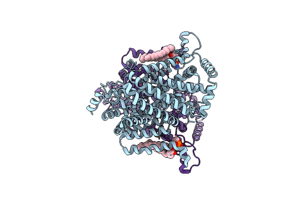
Organism: Klebsiella pneumoniae, Homo sapiens, Synthetic construct
Method: ELECTRON MICROSCOPY Release Date: 2025-03-12 Classification: STRUCTURAL PROTEIN/IMMUNE SYSTEM Ligands: CIT, PLM, NAG |
 |
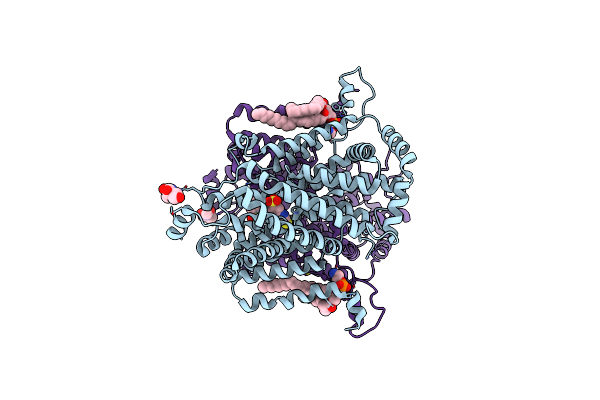
Organism: Klebsiella pneumoniae, Synthetic construct, Homo sapiens
Method: ELECTRON MICROSCOPY Release Date: 2025-03-12 Classification: STRUCTURAL PROTEIN/IMMUNE SYSTEM Ligands: CIT, PLM, NAG |
 |
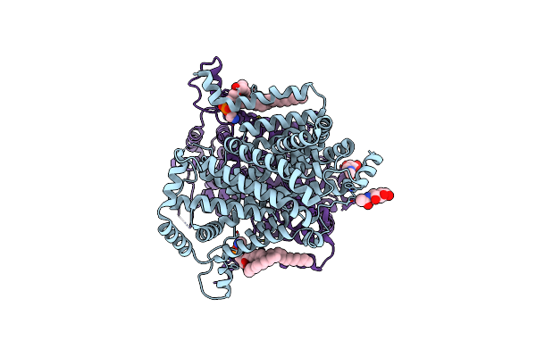
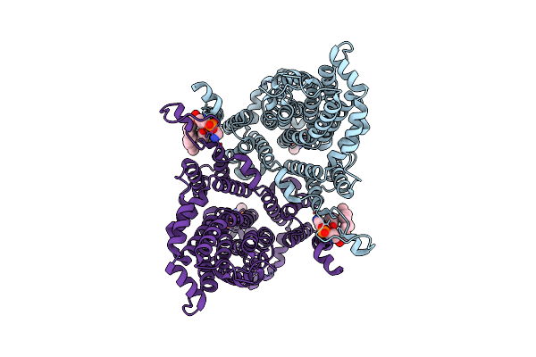
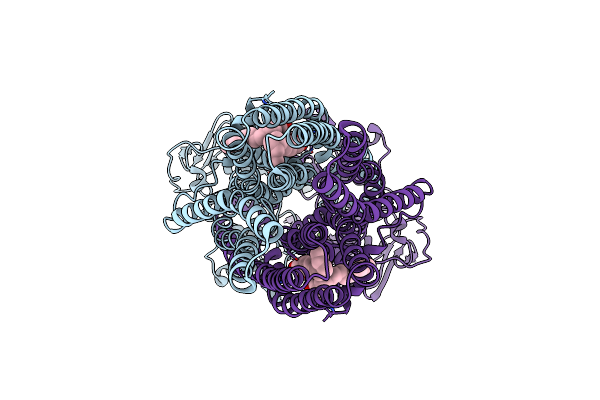
Cryo-Em Structure Of The G15C-R66C And T83C-T83C Diabody Complex (Cits-Diabody #7-Tlr3)
Organism: Klebsiella pneumoniae, Homo sapiens, Synthetic construct
Method: ELECTRON MICROSCOPY Release Date: 2025-03-12 Classification: STRUCTURAL PROTEIN/IMMUNE SYSTEM Ligands: CIT, PLM, NAG |
 |
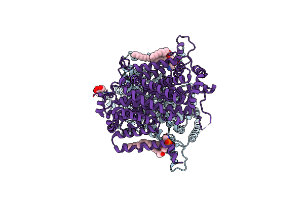
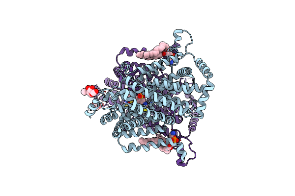
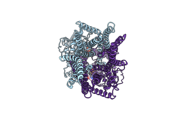
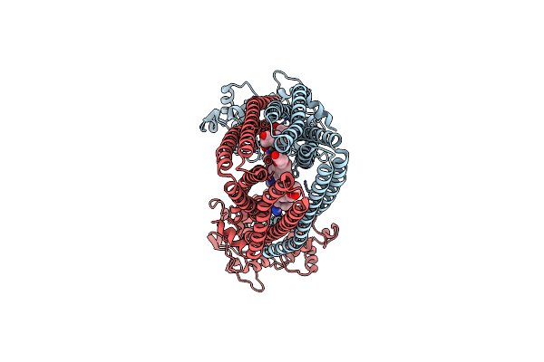
Cryo-Em Structure Of The Klebsiella Pneumoniae Cits (Citrate-Bound Occluded State)
Organism: Klebsiella pneumoniae
Method: ELECTRON MICROSCOPY Release Date: 2025-03-12 Classification: MEMBRANE PROTEIN Ligands: CIT, PLM |
 |
Organism: Drosophila melanogaster
Method: ELECTRON MICROSCOPY Release Date: 2025-01-29 Classification: MEMBRANE PROTEIN Ligands: PTY, NAG |
 |
Organism: Drosophila melanogaster
Method: ELECTRON MICROSCOPY Release Date: 2025-01-29 Classification: MEMBRANE PROTEIN Ligands: PTY, NAG |
 |
Organism: Drosophila melanogaster
Method: ELECTRON MICROSCOPY Release Date: 2025-01-29 Classification: MEMBRANE PROTEIN Ligands: PTY |
 |
Organism: Drosophila melanogaster
Method: ELECTRON MICROSCOPY Release Date: 2025-01-29 Classification: MEMBRANE PROTEIN Ligands: 4DS, PTY, NAG |
 |
Organism: Drosophila melanogaster
Method: ELECTRON MICROSCOPY Release Date: 2025-01-29 Classification: MEMBRANE PROTEIN Ligands: PTY, NAG, 4DS |
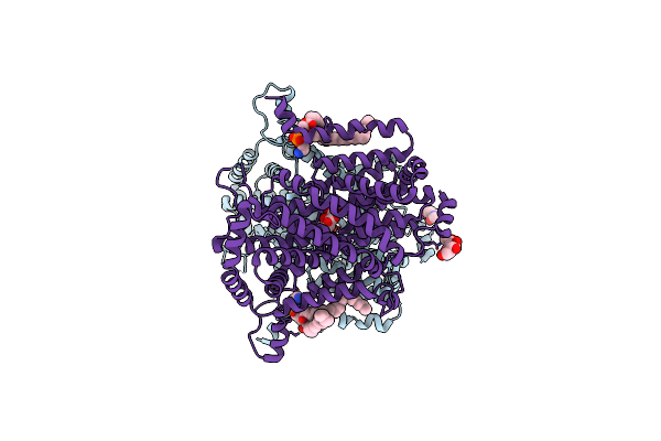 |
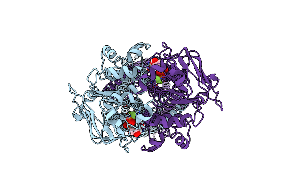
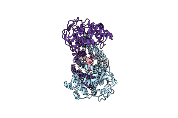
Cryo-Em Structure Of The Drosophila Indy (Citrate-Bound Inward-Occluded, Ph 6)
Organism: Drosophila melanogaster
Method: ELECTRON MICROSCOPY Release Date: 2025-01-29 Classification: MEMBRANE PROTEIN Ligands: FLC, NAG, PTY |
 |
Organism: Drosophila melanogaster
Method: ELECTRON MICROSCOPY Release Date: 2025-01-29 Classification: MEMBRANE PROTEIN Ligands: PTY, NAG |
 |
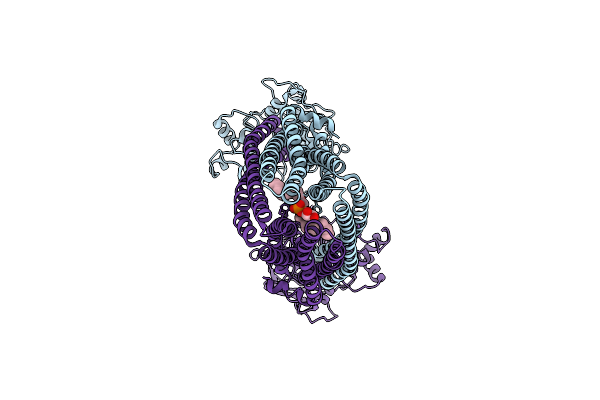
Crystal Structure Of The Gamma-Carbonic Anhydrase From The Polyextremophilic Bacterium Aeribacillus Pallidus
Organism: Aeribacillus pallidus
Method: X-RAY DIFFRACTION Resolution:1.70 Å Release Date: 2024-12-11 Classification: METAL BINDING PROTEIN Ligands: ZN, SO4 |
 |
Organism: Homo sapiens
Method: ELECTRON MICROSCOPY Release Date: 2024-06-19 Classification: MEMBRANE PROTEIN Ligands: GSH, CD |
 |
Cryo-Em Structure Of The Human Abcb6 In Complex With Cd(Ii):Phytochelatin 2
Organism: Homo sapiens, Tracheophyta
Method: ELECTRON MICROSCOPY Release Date: 2024-06-19 Classification: MEMBRANE PROTEIN Ligands: CD |
 |
Organism: Homo sapiens
Method: ELECTRON MICROSCOPY Release Date: 2023-10-04 Classification: TRANSPORT PROTEIN Ligands: PEV, AOV, MG |
 |
Organism: Homo sapiens
Method: ELECTRON MICROSCOPY Release Date: 2023-10-04 Classification: TRANSPORT PROTEIN Ligands: ADP, VO4, MG |
 |
Organism: Mus musculus
Method: ELECTRON MICROSCOPY Release Date: 2022-10-19 Classification: MEMBRANE PROTEIN Ligands: ADP, BEF, MG |
 |
Organism: Mus musculus
Method: ELECTRON MICROSCOPY Release Date: 2022-10-19 Classification: MEMBRANE PROTEIN Ligands: PGT |
 |
Organism: Mus musculus
Method: ELECTRON MICROSCOPY Release Date: 2022-10-19 Classification: MEMBRANE PROTEIN Ligands: Y01 |
 |
Organism: Homo sapiens
Method: ELECTRON MICROSCOPY Release Date: 2022-06-22 Classification: MEMBRANE PROTEIN Ligands: Y01, HT9 |