
Search Count: 37
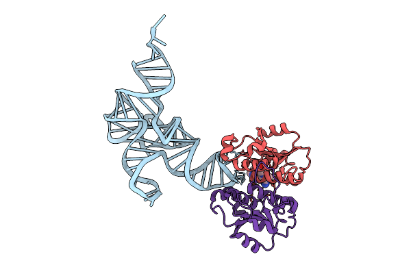 |
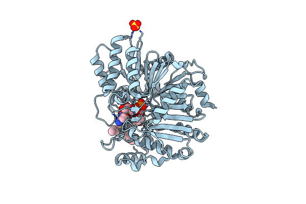
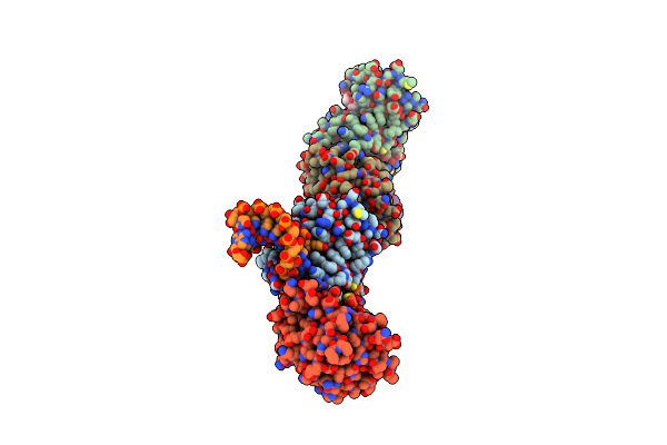
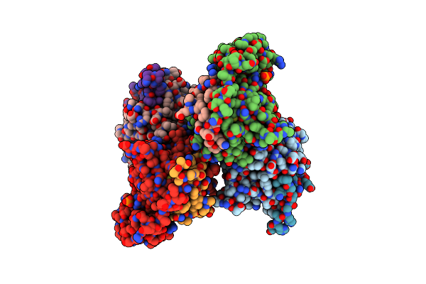
Crystal Structure Of B. Subtilis Cspr Complexed With Sinefungin And Cellularly Expressed Trna Leu
Organism: Bacillus subtilis subsp. subtilis str. 168
Method: X-RAY DIFFRACTION Release Date: 2025-07-23 Classification: TRANSFERASE Ligands: SFG |
 |
Organism: Escherichia coli k-12
Method: X-RAY DIFFRACTION Release Date: 2025-07-09 Classification: RNA |
 |
Organism: Bacillus subtilis subsp. subtilis str. 168
Method: X-RAY DIFFRACTION Release Date: 2025-07-09 Classification: RNA Ligands: SPM |
 |
Organism: Escherichia coli str. k-12 substr. mg1655
Method: X-RAY DIFFRACTION Resolution:2.83 Å Release Date: 2024-11-20 Classification: TRANSFERASE Ligands: SO4 |
 |
Crystal Structure Of E. Coli Phosphatidylserine Synthase Complexed With 16:0/16:0 Cdp-Dg
Organism: Escherichia coli str. k-12 substr. mg1655
Method: X-RAY DIFFRACTION Resolution:2.44 Å Release Date: 2024-11-20 Classification: TRANSFERASE Ligands: A1LZV, SO4 |
 |
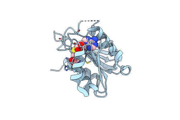
Organism: Bacillus subtilis subsp. subtilis str. 168
Method: X-RAY DIFFRACTION Release Date: 2024-09-11 Classification: TRANSFERASE Ligands: SO4, SAH, GOL |
 |
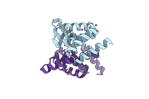
Organism: Homo sapiens
Method: X-RAY DIFFRACTION Release Date: 2024-05-15 Classification: APOPTOSIS Ligands: BME, SO4 |
 |
Organism: Homo sapiens
Method: X-RAY DIFFRACTION Release Date: 2023-08-02 Classification: APOPTOSIS Ligands: ZN |
 |
Organism: Homo sapiens
Method: X-RAY DIFFRACTION Release Date: 2023-07-05 Classification: APOPTOSIS Ligands: BME, PEG, NA |
 |
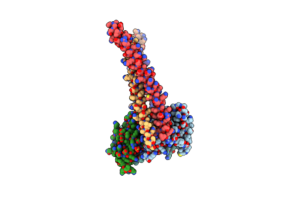
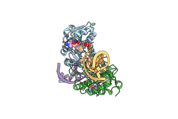
Crystal Structure Of Mnmm From B. Subtilis Complexed With Gln-Ttg Anti-Codon Stem Loop And Sam (2.90 A)
Organism: Bacillus subtilis subsp. subtilis str. 168
Method: X-RAY DIFFRACTION Resolution:2.90 Å Release Date: 2023-01-25 Classification: TRANSFERASE/RNA Ligands: SAM |
 |
Organism: Bacillus subtilis subsp. subtilis str. 168
Method: X-RAY DIFFRACTION Resolution:1.17 Å Release Date: 2023-01-25 Classification: TRANSFERASE Ligands: SAH |
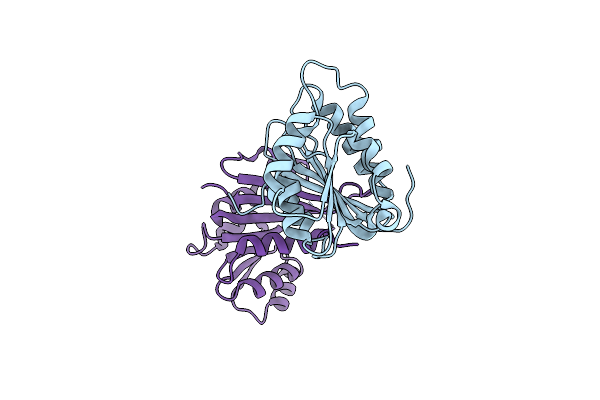 |
Organism: Staphylococcus aureus subsp. aureus nctc 8325
Method: X-RAY DIFFRACTION Resolution:1.44 Å Release Date: 2023-01-25 Classification: TRANSFERASE |
 |
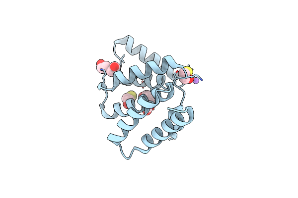
Crystal Structure Of Mnmm From S. Aureus Complexed With Sam And Trna Anti-Codon Stem Loop (Asl) (1.55 A)
Organism: Staphylococcus aureus subsp. aureus nctc 8325
Method: X-RAY DIFFRACTION Resolution:1.55 Å Release Date: 2023-01-25 Classification: TRANSFERASE/RNA Ligands: SAM, NA |
 |
Organism: Staphylococcus aureus subsp. aureus nctc 8325
Method: X-RAY DIFFRACTION Resolution:2.04 Å Release Date: 2023-01-25 Classification: TRANSFERASE Ligands: SAM |
 |
Organism: Staphylococcus aureus subsp. aureus nctc 8325
Method: X-RAY DIFFRACTION Release Date: 2023-01-18 Classification: TRANSFERASE Ligands: SAH |
 |
Organism: Escherichia coli k-12
Method: X-RAY DIFFRACTION Resolution:1.90 Å Release Date: 2021-03-24 Classification: LYASE Ligands: LMT, NA |
 |
Organism: Escherichia coli k-12
Method: X-RAY DIFFRACTION Resolution:2.63 Å Release Date: 2021-03-24 Classification: LYASE |
 |
Organism: Escherichia coli k-12
Method: X-RAY DIFFRACTION Resolution:2.12 Å Release Date: 2021-03-24 Classification: LYASE Ligands: LMT, G8C |
 |
Organism: Escherichia coli k-12
Method: X-RAY DIFFRACTION Resolution:2.70 Å Release Date: 2021-03-24 Classification: LYASE Ligands: PO4, PEX |
 |
Application Of Anti-Helix Antibodies In Protein Structure Determination (8189-3Lrh)
Organism: Staphylococcus aureus, Homo sapiens
Method: X-RAY DIFFRACTION Resolution:1.80 Å Release Date: 2019-08-14 Classification: STRUCTURAL PROTEIN |