
Search Count: 31
 |
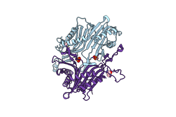
Organism: Vibrio campbellii
Method: X-RAY DIFFRACTION Resolution:2.00 Å Release Date: 2023-09-27 Classification: TOXIN Ligands: SO4 |
 |
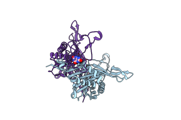
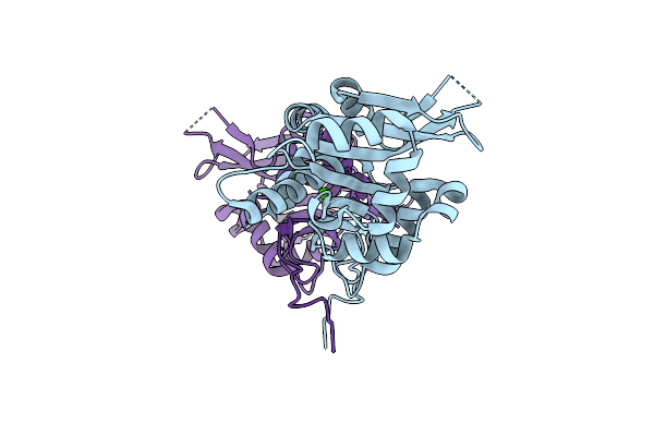
Organism: Vibrio campbellii
Method: ELECTRON MICROSCOPY Release Date: 2023-09-27 Classification: TOXIN Ligands: K, CA |
 |
Organism: Staphylococcus aureus
Method: X-RAY DIFFRACTION Resolution:1.87 Å Release Date: 2022-05-18 Classification: DNA BINDING PROTEIN |
 |
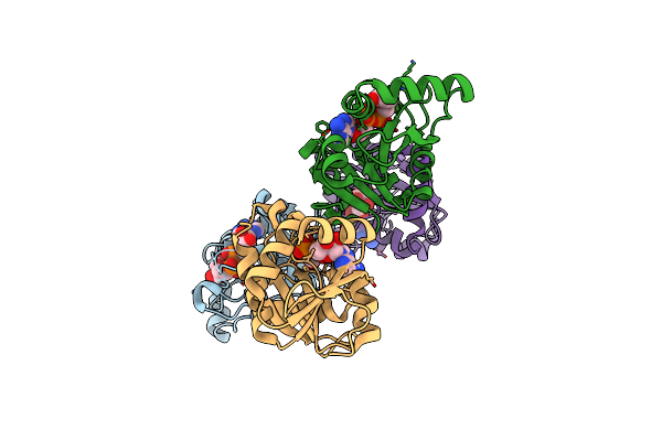
Organism: Staphylococcus aureus, Staphylococcus aureus subsp. aureus mu50
Method: X-RAY DIFFRACTION Resolution:2.00 Å Release Date: 2022-05-18 Classification: DNA BINDING PROTEIN/DNA Ligands: MG |
 |
Organism: Staphylococcus aureus (strain mu50 / atcc 700699)
Method: X-RAY DIFFRACTION Resolution:2.77 Å Release Date: 2022-05-18 Classification: DNA BINDING PROTEIN Ligands: BEF, MG |
 |
Organism: Thermobispora bispora (strain atcc 19993 / dsm 43833 / cbs 139.67 / jcm 10125 / nbrc 14880 / r51)
Method: X-RAY DIFFRACTION Resolution:1.60 Å Release Date: 2022-05-04 Classification: OXIDOREDUCTASE Ligands: SO4, DMS |
 |
Organism: Thermobispora bispora (strain atcc 19993 / dsm 43833 / cbs 139.67 / jcm 10125 / nbrc 14880 / r51)
Method: X-RAY DIFFRACTION Resolution:2.16 Å Release Date: 2022-05-04 Classification: OXIDOREDUCTASE Ligands: URC |
 |
Crystal Structure Of C-Terminal Dna-Binding Domain Of Escherichia Coli Ompr
Organism: Escherichia coli
Method: X-RAY DIFFRACTION Resolution:3.56 Å Release Date: 2020-12-23 Classification: DNA BINDING PROTEIN |
 |
Crystal Structure Of C-Terminal Dna-Binding Domain Of Escherichia Coli Ompr As A Domain-Swapped Dimer
Organism: Escherichia coli
Method: X-RAY DIFFRACTION Resolution:2.41 Å Release Date: 2020-12-23 Classification: DNA BINDING PROTEIN Ligands: BTB, SO4, GOL |
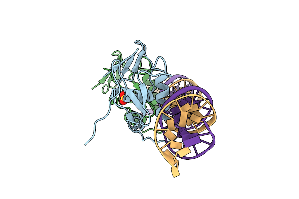 |
Crystal Structure Of C-Terminal Dna-Binding Domain Of Escherichia Coli Ompr In Complex With F1-Dna
Organism: Escherichia coli
Method: X-RAY DIFFRACTION Resolution:2.93 Å Release Date: 2020-12-23 Classification: DNA BINDING PROTEIN Ligands: SO4 |
 |
Organism: Saccharomyces cerevisiae s288c
Method: X-RAY DIFFRACTION Resolution:2.36 Å Release Date: 2020-12-09 Classification: HYDROLASE Ligands: GOL |
 |
Organism: Saccharomyces cerevisiae s288c
Method: X-RAY DIFFRACTION Resolution:1.78 Å Release Date: 2020-12-09 Classification: HYDROLASE Ligands: APR |
 |
Organism: Saccharomyces cerevisiae s288c
Method: X-RAY DIFFRACTION Resolution:1.87 Å Release Date: 2020-12-09 Classification: HYDROLASE Ligands: APR, CXS |
 |
Organism: Saccharomyces cerevisiae s288c
Method: X-RAY DIFFRACTION Resolution:1.57 Å Release Date: 2020-12-09 Classification: HYDROLASE Ligands: ACT, APR |
 |
Organism: Saccharomyces cerevisiae s288c
Method: X-RAY DIFFRACTION Resolution:3.12 Å Release Date: 2020-12-09 Classification: HYDROLASE Ligands: APR |
 |
Organism: Saccharomyces cerevisiae (strain atcc 204508 / s288c)
Method: X-RAY DIFFRACTION Resolution:1.83 Å Release Date: 2020-12-09 Classification: CHAPERONE Ligands: CA |
 |
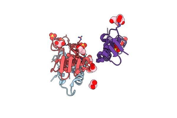
Organism: Severe acute respiratory syndrome coronavirus 2
Method: X-RAY DIFFRACTION Resolution:3.83 Å Release Date: 2020-11-11 Classification: HYDROLASE Ligands: APR |
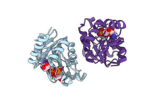 |
Organism: Severe acute respiratory syndrome coronavirus 2
Method: X-RAY DIFFRACTION Resolution:2.64 Å Release Date: 2020-11-11 Classification: VIRAL PROTEIN Ligands: APR |
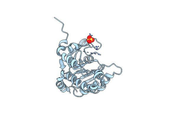 |
Crystal Structure Of Poly(Adp-Ribose) Glycohydrolase (Parg) From Deinococcus Radiodurans In Apo Form
Organism: Deinococcus radiodurans (strain atcc 13939 / dsm 20539 / jcm 16871 / lmg 4051 / nbrc 15346 / ncimb 9279 / r1 / vkm b-1422)
Method: X-RAY DIFFRACTION Resolution:1.55 Å Release Date: 2019-02-27 Classification: HYDROLASE Ligands: SO4 |
 |
Crystal Structure Of Poly(Adp-Ribose) Glycohydrolase (Parg) From Deinococcus Radiodurans In Complex With Adp-Ribose (P21)
Organism: Deinococcus radiodurans (strain atcc 13939 / dsm 20539 / jcm 16871 / lmg 4051 / nbrc 15346 / ncimb 9279 / r1 / vkm b-1422)
Method: X-RAY DIFFRACTION Resolution:1.97 Å Release Date: 2019-02-27 Classification: HYDROLASE Ligands: AR6 |