
Search Count: 62
 |
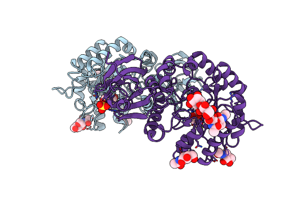
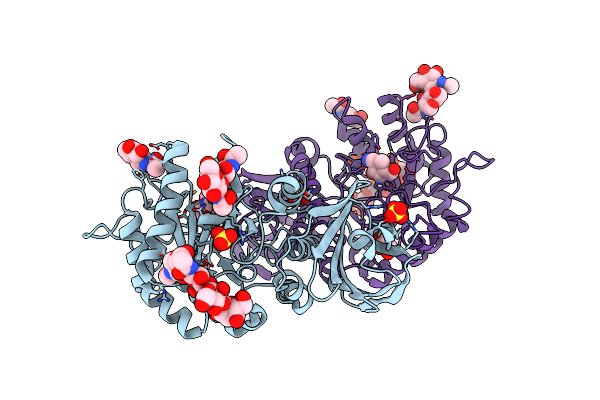
Crystal Structure Of Polyketide Synthase (Pks) Thioreductase Domain From Streptomyces Coelicolor
Organism: Streptomyces coelicolor
Method: X-RAY DIFFRACTION Resolution:1.84 Å Release Date: 2024-11-27 Classification: BIOSYNTHETIC PROTEIN Ligands: NDP, GOL, BTB |
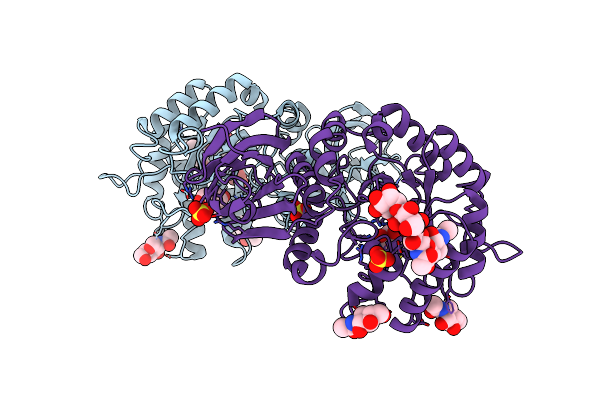 |
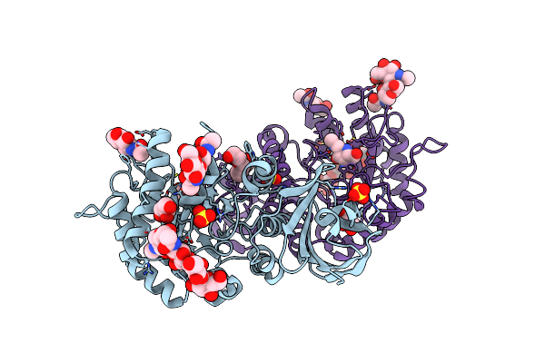
Crystal Structure Of Human Lysosomal Alpha-Galactosidase A In Complex With (2R,3R,4S,5R)-2-(Aminomethyl)-5-(Hydroxymethyl)Pyrrolidine-3,4-Diol
Organism: Homo sapiens
Method: X-RAY DIFFRACTION Resolution:2.61 Å Release Date: 2024-04-10 Classification: HYDROLASE Ligands: NAG, VLN, SO4 |
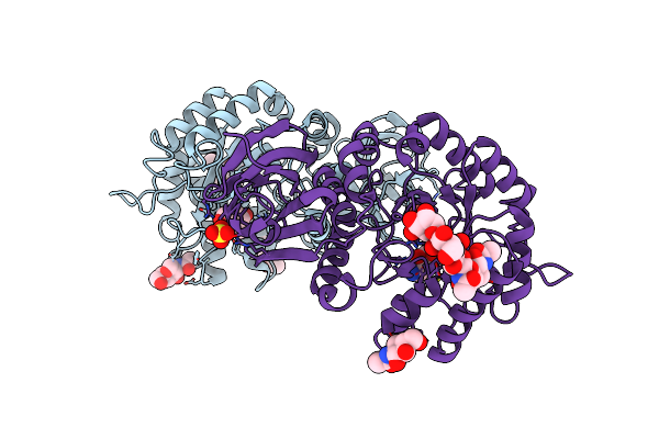 |
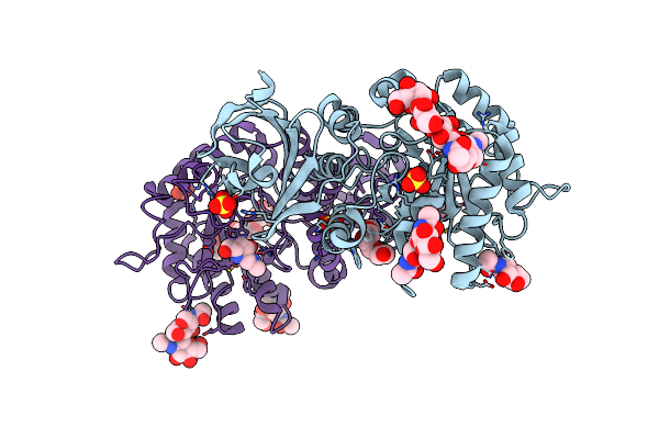
Crystal Structure Of Human Lysosomal Alpha-Galactosidase A In Complex With (2R,3R,4S,5R)-2-(Aminomethyl)-5-(Hydroxymethyl)Pyrrolidine-3,4-Diol
Organism: Homo sapiens
Method: X-RAY DIFFRACTION Resolution:2.20 Å Release Date: 2024-04-10 Classification: HYDROLASE Ligands: NAG, VLN, SO4 |
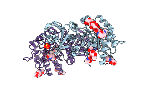 |
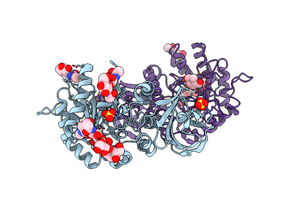
Crystal Structure Of Human Lysosomal Alpha-Galactosidase A In Complex With (2R,3S,4R,5R)-2,5-Bis(Hydroxymethyl)Pyrrolidine-3,4-Diol
Organism: Homo sapiens
Method: X-RAY DIFFRACTION Resolution:1.98 Å Release Date: 2024-04-10 Classification: HYDROLASE Ligands: NAG, VMF, SO4 |
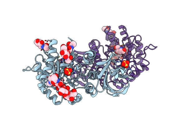 |
Crystal Structure Of Human Lysosomal Alpha-Galactosidase A In Complex With (2R,3S,4R,5R)-2,5-Bis(Hydroxymethyl)Pyrrolidine-3,4-Diol
Organism: Homo sapiens
Method: X-RAY DIFFRACTION Resolution:2.32 Å Release Date: 2024-04-10 Classification: HYDROLASE Ligands: NAG, VMF, SO4 |
 |
Crystal Structure Of Human Lysosomal Alpha-Galactosidase A In Complex With (2R,3S,4R)-2-(Hydroxymethyl)-1-Methylpyrrolidine-3,4-Diol
Organism: Homo sapiens
Method: X-RAY DIFFRACTION Resolution:2.28 Å Release Date: 2024-04-10 Classification: HYDROLASE Ligands: NAG, VMO, SO4 |
 |
Crystal Structure Of Human Lysosomal Alpha-Galactosidase A In Complex With (2R,3S,4R,5R)-2-(Hydroxymethyl)-5-((Methylamino)Methyl)Pyrrolidine-3,4-Diol
Organism: Homo sapiens
Method: X-RAY DIFFRACTION Resolution:2.12 Å Release Date: 2024-04-10 Classification: HYDROLASE Ligands: NAG, VN0, SO4, P6G, GOL |
 |
Crystal Structure Of Human Lysosomal Alpha-Galactosidase A In Complex With (2R,3R,4S,5R)-2-((Dimethylamino)Methyl)-5-(Hydroxymethyl)Pyrrolidine-3,4-Diol
Organism: Homo sapiens
Method: X-RAY DIFFRACTION Resolution:2.01 Å Release Date: 2024-04-10 Classification: HYDROLASE Ligands: NAG, VNB, SO4, P6G |
 |
Crystal Structure Of Human Lysosomal Alpha-Galactosidase A In Complex With (2R,3S,4R,5R)-2,5-Bis(Hydroxymethyl)-1-Methylpyrrolidine-3,4-Diol
Organism: Homo sapiens
Method: X-RAY DIFFRACTION Resolution:2.00 Å Release Date: 2024-04-10 Classification: HYDROLASE Ligands: NAG, VNQ, SO4 |
 |
Crystal Structure Of Human Lysosomal Alpha-Galactosidase A In Complex With (2R,3R,4S,5R)-2-(Aminomethyl)-5-(Hydroxymethyl)-1-Methylpyrrolidine-3,4-Diol
Organism: Homo sapiens
Method: X-RAY DIFFRACTION Resolution:2.00 Å Release Date: 2024-04-10 Classification: HYDROLASE Ligands: NAG, VO0, SO4, GOL |
 |
Organism: Vibrio campbellii
Method: X-RAY DIFFRACTION Resolution:2.00 Å Release Date: 2023-09-27 Classification: TOXIN Ligands: SO4 |
 |
Organism: Vibrio campbellii
Method: ELECTRON MICROSCOPY Release Date: 2023-09-27 Classification: TOXIN Ligands: K, CA |
 |
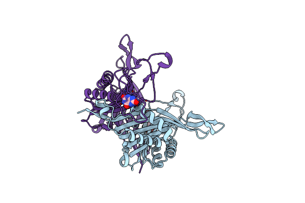
Parathyroid Hormone 1 Receptor Extracellular Domain Complexed With A Peptide Ligand Containing Beta-3-Homotryptophan
Organism: Homo sapiens
Method: X-RAY DIFFRACTION Resolution:2.00 Å Release Date: 2023-06-14 Classification: SIGNALING PROTEIN Ligands: EDO, ZN |
 |
Parathyroid Hormone 1 Receptor Extracellular Domain Complexed With A Peptide Ligand Containing (2-Naphthyl)-Beta-3-Homoalanine
Organism: Homo sapiens
Method: X-RAY DIFFRACTION Resolution:2.02 Å Release Date: 2023-06-14 Classification: SIGNALING PROTEIN Ligands: EDO, ZN |
 |
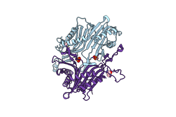
Crystal Structure Of Canine Coronavirus Main Protease In Complex With Gc376
Organism: Canine coronavirus
Method: X-RAY DIFFRACTION Resolution:2.75 Å Release Date: 2023-05-31 Classification: VIRAL PROTEIN/INHIBITOR Ligands: K36 |
 |
Organism: Staphylococcus aureus
Method: X-RAY DIFFRACTION Resolution:1.87 Å Release Date: 2022-05-18 Classification: DNA BINDING PROTEIN |
 |
Organism: Staphylococcus aureus, Staphylococcus aureus subsp. aureus mu50
Method: X-RAY DIFFRACTION Resolution:2.00 Å Release Date: 2022-05-18 Classification: DNA BINDING PROTEIN/DNA Ligands: MG |
 |
Organism: Staphylococcus aureus (strain mu50 / atcc 700699)
Method: X-RAY DIFFRACTION Resolution:2.77 Å Release Date: 2022-05-18 Classification: DNA BINDING PROTEIN Ligands: BEF, MG |
 |
Organism: Thermobispora bispora (strain atcc 19993 / dsm 43833 / cbs 139.67 / jcm 10125 / nbrc 14880 / r51)
Method: X-RAY DIFFRACTION Resolution:1.60 Å Release Date: 2022-05-04 Classification: OXIDOREDUCTASE Ligands: SO4, DMS |
 |
Organism: Thermobispora bispora (strain atcc 19993 / dsm 43833 / cbs 139.67 / jcm 10125 / nbrc 14880 / r51)
Method: X-RAY DIFFRACTION Resolution:2.16 Å Release Date: 2022-05-04 Classification: OXIDOREDUCTASE Ligands: URC |