
Search Count: 32
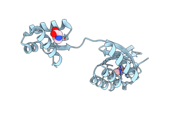 |
Organism: Thermotoga maritima
Method: X-RAY DIFFRACTION Resolution:2.30 Å Release Date: 2025-01-22 Classification: DNA BINDING PROTEIN Ligands: FE2, NIO, PRO |
 |
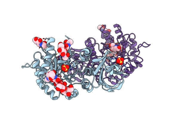
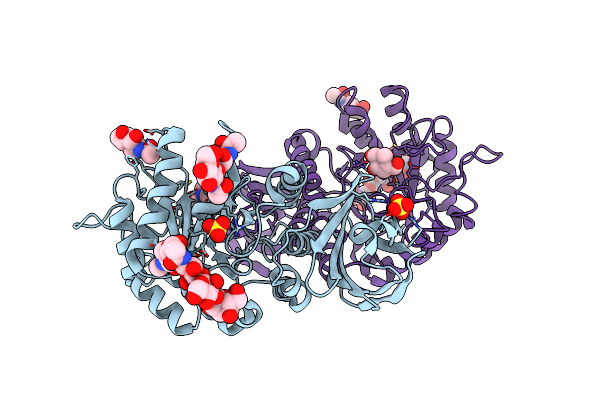
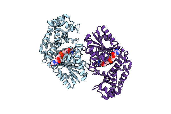
Crystal Structure Of Human Lysosomal Alpha-Galactosidase A In Complex With (2R,3R,4S,5R)-2-(Aminomethyl)-5-(Hydroxymethyl)Pyrrolidine-3,4-Diol
Organism: Homo sapiens
Method: X-RAY DIFFRACTION Resolution:2.61 Å Release Date: 2024-04-10 Classification: HYDROLASE Ligands: NAG, VLN, SO4 |
 |
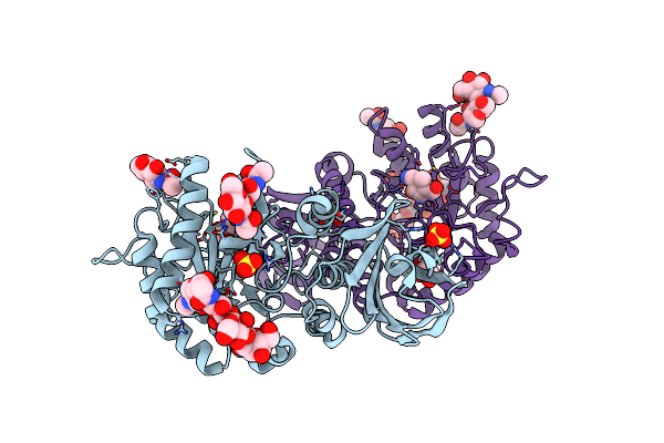
Crystal Structure Of Human Lysosomal Alpha-Galactosidase A In Complex With (2R,3R,4S,5R)-2-(Aminomethyl)-5-(Hydroxymethyl)Pyrrolidine-3,4-Diol
Organism: Homo sapiens
Method: X-RAY DIFFRACTION Resolution:2.20 Å Release Date: 2024-04-10 Classification: HYDROLASE Ligands: NAG, VLN, SO4 |
 |
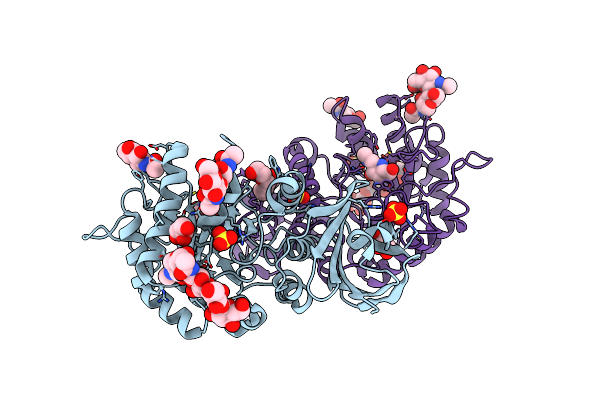
Crystal Structure Of Human Lysosomal Alpha-Galactosidase A In Complex With (2R,3S,4R,5R)-2,5-Bis(Hydroxymethyl)Pyrrolidine-3,4-Diol
Organism: Homo sapiens
Method: X-RAY DIFFRACTION Resolution:1.98 Å Release Date: 2024-04-10 Classification: HYDROLASE Ligands: NAG, VMF, SO4 |
 |
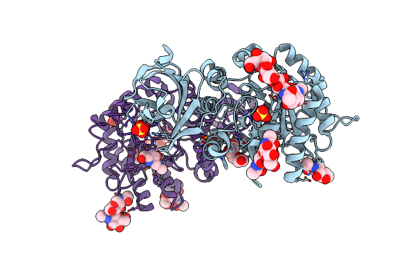
Crystal Structure Of Human Lysosomal Alpha-Galactosidase A In Complex With (2R,3S,4R,5R)-2,5-Bis(Hydroxymethyl)Pyrrolidine-3,4-Diol
Organism: Homo sapiens
Method: X-RAY DIFFRACTION Resolution:2.32 Å Release Date: 2024-04-10 Classification: HYDROLASE Ligands: NAG, VMF, SO4 |
 |
Crystal Structure Of Human Lysosomal Alpha-Galactosidase A In Complex With (2R,3S,4R)-2-(Hydroxymethyl)-1-Methylpyrrolidine-3,4-Diol
Organism: Homo sapiens
Method: X-RAY DIFFRACTION Resolution:2.28 Å Release Date: 2024-04-10 Classification: HYDROLASE Ligands: NAG, VMO, SO4 |
 |
Crystal Structure Of Human Lysosomal Alpha-Galactosidase A In Complex With (2R,3S,4R,5R)-2-(Hydroxymethyl)-5-((Methylamino)Methyl)Pyrrolidine-3,4-Diol
Organism: Homo sapiens
Method: X-RAY DIFFRACTION Resolution:2.12 Å Release Date: 2024-04-10 Classification: HYDROLASE Ligands: NAG, VN0, SO4, P6G, GOL |
 |
Crystal Structure Of Human Lysosomal Alpha-Galactosidase A In Complex With (2R,3R,4S,5R)-2-((Dimethylamino)Methyl)-5-(Hydroxymethyl)Pyrrolidine-3,4-Diol
Organism: Homo sapiens
Method: X-RAY DIFFRACTION Resolution:2.01 Å Release Date: 2024-04-10 Classification: HYDROLASE Ligands: NAG, VNB, SO4, P6G |
 |
Crystal Structure Of Human Lysosomal Alpha-Galactosidase A In Complex With (2R,3S,4R,5R)-2,5-Bis(Hydroxymethyl)-1-Methylpyrrolidine-3,4-Diol
Organism: Homo sapiens
Method: X-RAY DIFFRACTION Resolution:2.00 Å Release Date: 2024-04-10 Classification: HYDROLASE Ligands: NAG, VNQ, SO4 |
 |
Crystal Structure Of Human Lysosomal Alpha-Galactosidase A In Complex With (2R,3R,4S,5R)-2-(Aminomethyl)-5-(Hydroxymethyl)-1-Methylpyrrolidine-3,4-Diol
Organism: Homo sapiens
Method: X-RAY DIFFRACTION Resolution:2.00 Å Release Date: 2024-04-10 Classification: HYDROLASE Ligands: NAG, VO0, SO4, GOL |
 |
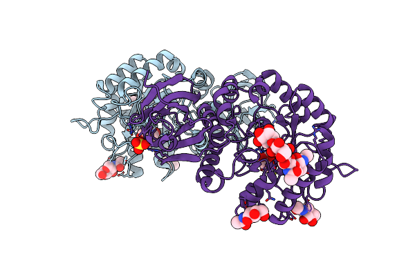
Crystal Structure Of Shikimate Dehydrogenase (Aroe) Clinical Variant V2356 From Helicobacter Pylori In Complex With Shikimate
Organism: Helicobacter pylori
Method: X-RAY DIFFRACTION Resolution:2.85 Å Release Date: 2013-01-30 Classification: OXIDOREDUCTASE Ligands: SKM |
 |
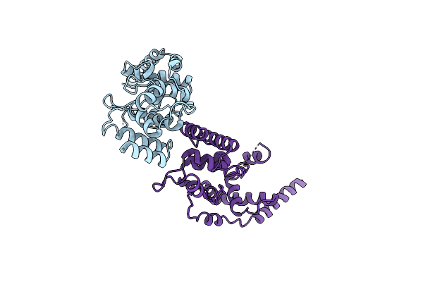
Crystal Structure Of Staphylococcus Aureus Membrane-Bound Transglycosylase: Apoenzyme
Organism: Staphylococcus aureus
Method: X-RAY DIFFRACTION Resolution:2.52 Å Release Date: 2012-04-18 Classification: TRANSFERASE Ligands: MG |
 |
Crystal Structure Of Staphylococcus Aureus Membrane-Bound Transglycosylase In Complex With Moenomycin
Organism: Staphylococcus aureus
Method: X-RAY DIFFRACTION Resolution:3.69 Å Release Date: 2012-04-18 Classification: TRANSFERASE Ligands: M0E |
 |
Crystal Structure Of Staphylococcus Aureus Membrane-Bound Transglycosylase In Complex With Nbd-Lipid Ii
Organism: Staphylococcus aureus
Method: X-RAY DIFFRACTION Resolution:3.20 Å Release Date: 2012-04-18 Classification: TRANSFERASE |
 |
Crystal Structure Of Staphylococcus Aureus Membrane-Bound Transglycosylase In Complex With A Lipid Ii Analog
Organism: Staphylococcus aureus
Method: X-RAY DIFFRACTION Resolution:2.30 Å Release Date: 2012-04-18 Classification: TRANSFERASE Ligands: LHI, MG |
 |
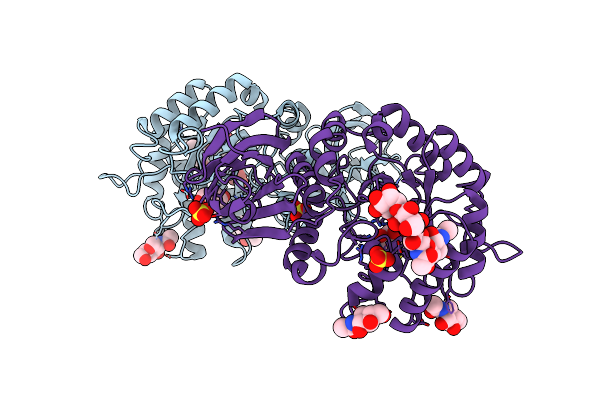
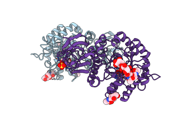
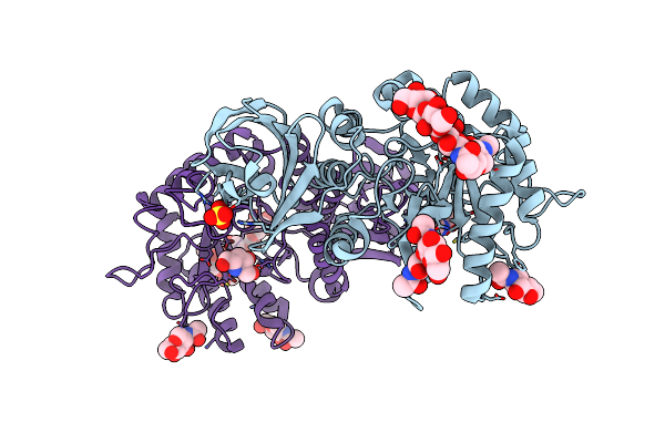
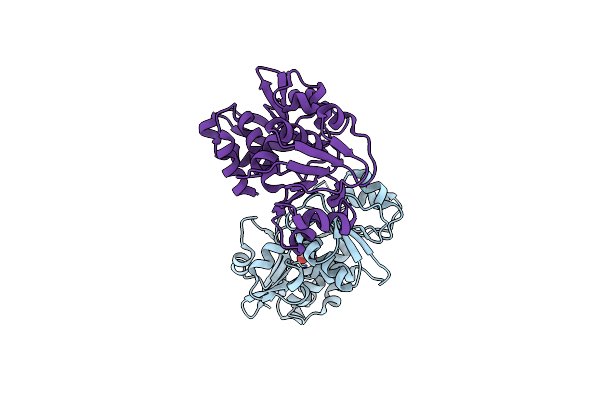
Crystal Structure Of The Full-Length Transglycosylase Pbp1B From Escherichia Coli
Organism: Escherichia coli
Method: X-RAY DIFFRACTION Resolution:2.16 Å Release Date: 2012-03-14 Classification: TRANSFERASE, HYDROLASE/ANTIBIOTIC Ligands: M0E |
 |
3-Dehydroquinate Synthase (Arob) From Mycobacterium Tuberculosis In Complex With Nad
Organism: Mycobacterium tuberculosis
Method: X-RAY DIFFRACTION Resolution:2.47 Å Release Date: 2012-01-18 Classification: LYASE Ligands: NAD |
 |
Crystal Structure Of The 3-Dehydroquinate Synthase (Arob) From Mycobacterium Tuberculosis
Organism: Mycobacterium tuberculosis
Method: X-RAY DIFFRACTION Resolution:2.07 Å Release Date: 2012-01-18 Classification: LYASE Ligands: ZN, CL |
 |
Crystal Structure Of The Shikimate 5-Dehydrogenase (Aroe) From Helicobacter Pylori
Organism: Helicobacter pylori
Method: X-RAY DIFFRACTION Resolution:1.57 Å Release Date: 2011-11-09 Classification: OXIDOREDUCTASE |
 |
Shikimate 5-Dehydrogenase (Aroe) From Helicobacter Pylori In Complex With Shikimate
Organism: Helicobacter pylori
Method: X-RAY DIFFRACTION Resolution:1.42 Å Release Date: 2011-11-09 Classification: OXIDOREDUCTASE Ligands: SKM |