
Search Count: 19
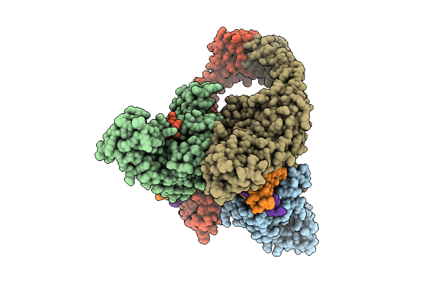 |
Organism: Escherichia phage t4, Homo sapiens
Method: ELECTRON MICROSCOPY Release Date: 2025-10-22 Classification: ISOMERASE Ligands: MG |
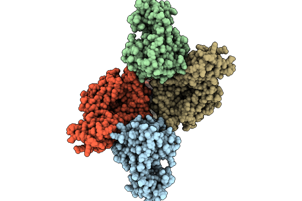 |
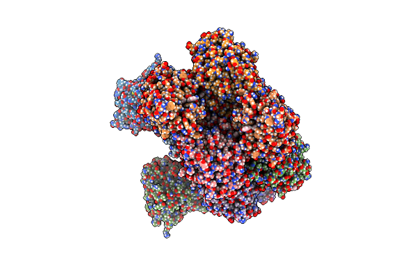
Organism: Escherichia phage t4, Dna molecule
Method: ELECTRON MICROSCOPY Release Date: 2025-10-01 Classification: ISOMERASE/DNA |
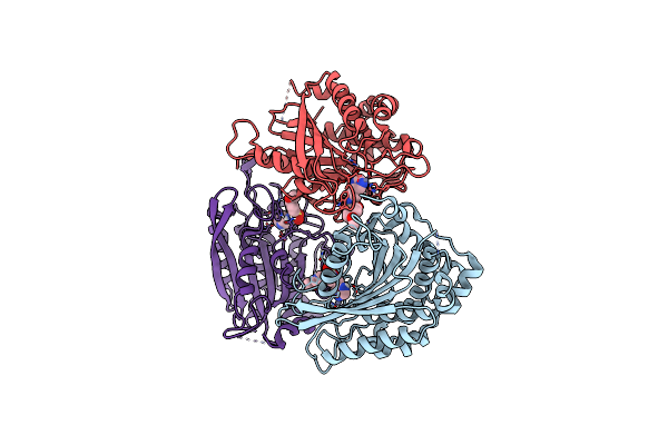 |
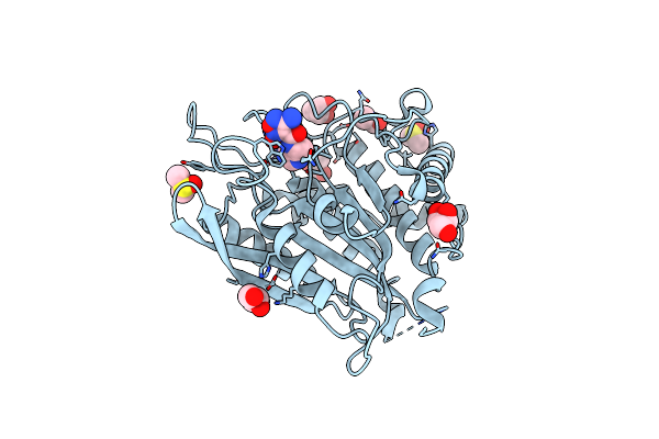
Crystal Structure Of Human Glutaminyl Cyclase In Complex With N-(2-(1H-Imidazol-5-Yl)Ethyl)-4-Methoxybenzenesulfonamide
Organism: Homo sapiens
Method: X-RAY DIFFRACTION Release Date: 2025-06-25 Classification: TRANSFERASE Ligands: ZN, A1EFY |
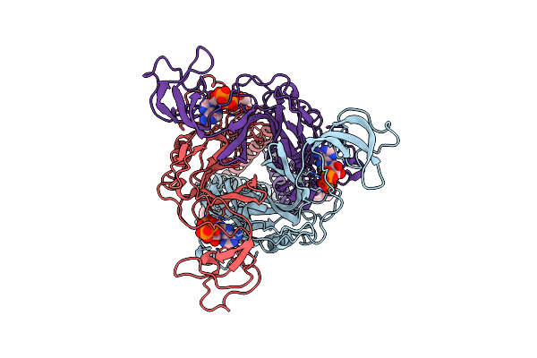 |
Organism: Homo sapiens
Method: ELECTRON MICROSCOPY Release Date: 2025-04-16 Classification: MEMBRANE PROTEIN Ligands: ATP, CLR |
 |
Organism: Homo sapiens
Method: ELECTRON MICROSCOPY Release Date: 2025-04-16 Classification: MEMBRANE PROTEIN Ligands: CLR, A1ALI |
 |
Crystal Structure Of Human Secretory Glutaminyl Cyclase In Complex With The Inhibitor N-(1H-Benzo[D]Imidazol-5-Yl)-1-Phenylmethanesulfonamide (Compound 5)
Organism: Homo sapiens
Method: X-RAY DIFFRACTION Resolution:2.37 Å Release Date: 2025-03-12 Classification: TRANSFERASE Ligands: ZN, A1D93, GOL, DMS, DMF, PEG |
 |
Crystal Structure Of Human Secretory Glutaminyl Cyclase In Complex With The Inhibitor 3-((2-(1H-Imidazol-5-Yl)Ethyl)Carbamoyl)-4-Amino-1,2,5-Oxadiazole 2-Oxide (Compound 13)
Organism: Homo sapiens
Method: X-RAY DIFFRACTION Resolution:2.96 Å Release Date: 2025-03-12 Classification: TRANSFERASE Ligands: ZN, A1D94, DMS, GOL, DMF |
 |
Organism: Homo sapiens
Method: X-RAY DIFFRACTION Resolution:1.91 Å Release Date: 2024-09-04 Classification: ANTIBIOTIC Ligands: MYR, A1AZR |
 |
Organism: Acinetobacter baumannii
Method: X-RAY DIFFRACTION Resolution:2.80 Å Release Date: 2024-09-04 Classification: ANTIBIOTIC Ligands: ANP, MVC, MG, A1AZS |
 |
Organism: Acinetobacter baumannii
Method: ELECTRON MICROSCOPY Release Date: 2024-04-24 Classification: ANTIMICROBIAL PROTEIN Ligands: ANP, ZQF |
 |
Organism: Homo sapiens
Method: X-RAY DIFFRACTION Resolution:2.10 Å Release Date: 2024-02-14 Classification: CELL CYCLE |
 |
Voltage-Gated Potassium Channel Kv3.1 With Novel Positive Modulator (9M)-9-{5-Chloro-6-[(3,3-Dimethyl-2,3-Dihydro-1-Benzofuran-4-Yl)Oxy]-4-Methylpyridin-3-Yl}-2-Methyl-7,9-Dihydro-8H-Purin-8-One (Compound 4)
Organism: Homo sapiens
Method: ELECTRON MICROSCOPY Release Date: 2023-10-25 Classification: TRANSPORT PROTEIN Ligands: ZN, X9T, POV, K |
 |
Organism: Homo sapiens
Method: ELECTRON MICROSCOPY Release Date: 2023-10-25 Classification: TRANSPORT PROTEIN Ligands: ZN, CLR, K |
 |
Organism: Synthetic construct
Method: X-RAY DIFFRACTION Resolution:1.88 Å Release Date: 2023-05-31 Classification: RNA |
 |
Crystal Structure Of Cug Repeat Rna Duplex Containing A-U Base Pair And U-U Mismatches
Organism: Synthetic construct
Method: X-RAY DIFFRACTION Resolution:1.58 Å Release Date: 2023-05-31 Classification: RNA |
 |
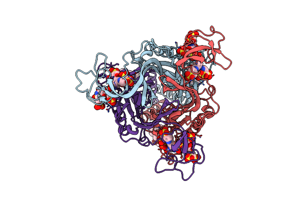
Organism: Homo sapiens, Severe acute respiratory syndrome coronavirus 2
Method: ELECTRON MICROSCOPY Release Date: 2021-04-21 Classification: VIRAL PROTEIN Ligands: NAG |
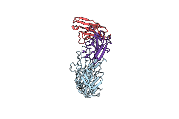 |
A Proof Of Concept For Neutralizing Antibody-Guided Vaccine Design Against Sars-Cov-2
Organism: Severe acute respiratory syndrome coronavirus 2, Homo sapiens
Method: ELECTRON MICROSCOPY Release Date: 2021-04-07 Classification: VIRAL PROTEIN |
 |
Crystal Structure Analysis Of Cruzain Bound To Vinyl Sulfone Derived Inhibitor (Wrr483)
Organism: Trypanosoma cruzi
Method: X-RAY DIFFRACTION Resolution:1.50 Å Release Date: 2010-10-20 Classification: HYDROLASE Ligands: 4MC, EDO, SO4 |
 |
Substrate Specificity Profiling And Identification Of A New Class Of Inhibitor For The Major Protease Of The Sars Coronavirus
Organism: Sars coronavirus
Method: X-RAY DIFFRACTION Resolution:1.80 Å Release Date: 2007-07-17 Classification: HYDROLASE Ligands: WR1 |