
Search Count: 3,026
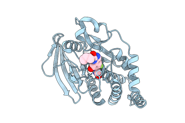 |
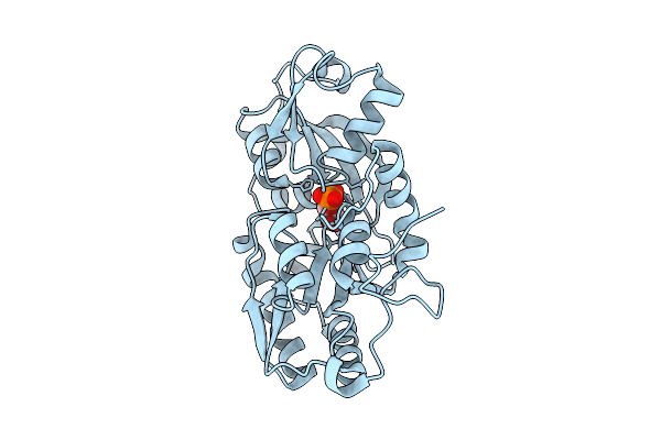
Organism: Homo sapiens
Method: X-RAY DIFFRACTION Release Date: 2025-12-17 Classification: IMMUNE SYSTEM Ligands: A1EQR |
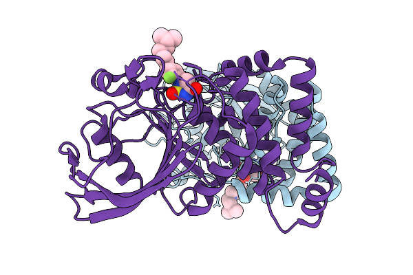 |
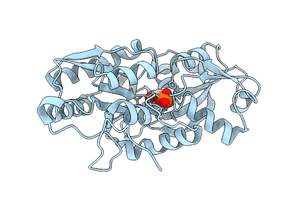
Organism: Homo sapiens
Method: X-RAY DIFFRACTION Release Date: 2025-12-17 Classification: IMMUNE SYSTEM Ligands: A1ETL |
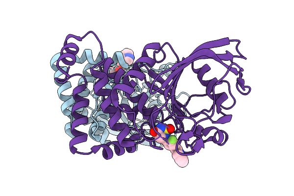 |
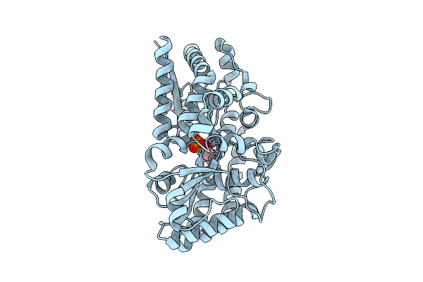
Organism: Homo sapiens
Method: X-RAY DIFFRACTION Release Date: 2025-12-17 Classification: IMMUNE SYSTEM Ligands: A1ETK |
 |
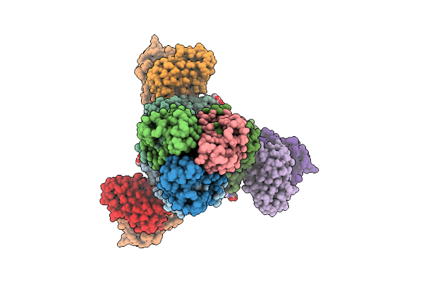
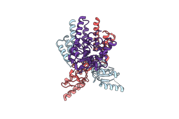
Cryo-Em Structure Of Conivaptan-Bound Human Vasopressin V2 Receptor Complex With Fab
Organism: Homo sapiens, Escherichia coli
Method: ELECTRON MICROSCOPY Release Date: 2025-12-10 Classification: MEMBRANE PROTEIN/IMMUNE SYSTEM Ligands: A1ECE |
 |
Cryo-Em Structure Of Tolvaptan-Bound Human Vasopressin V2 Receptor Complex With Fab
Organism: Homo sapiens, Escherichia coli
Method: ELECTRON MICROSCOPY Release Date: 2025-12-10 Classification: MEMBRANE PROTEIN/IMMUNE SYSTEM Ligands: A1ECF |
 |
Cryoem Structure Of H7 Hemagglutinin In Complex With A Human Neutralizing Antibody 6Y13
Organism: Influenza a virus (a/duck/chiba/25-51-14/2013(h7n1)), Homo sapiens
Method: ELECTRON MICROSCOPY Release Date: 2025-12-03 Classification: VIRAL PROTEIN Ligands: NAG |
 |
Organism: Escherichia coli
Method: ELECTRON MICROSCOPY Release Date: 2025-12-03 Classification: DNA BINDING PROTEIN Ligands: ZN |
 |
Organism: Marinobacter sp. dsm 11874
Method: X-RAY DIFFRACTION Release Date: 2025-11-26 Classification: TRANSPORT PROTEIN Ligands: 1GP |
 |
Organism: Marinobacter sp. dsm 11874
Method: X-RAY DIFFRACTION Release Date: 2025-11-26 Classification: TRANSPORT PROTEIN Ligands: G3P |
 |
Organism: Phaeobacter sp. med193
Method: X-RAY DIFFRACTION Release Date: 2025-11-26 Classification: TRANSPORT PROTEIN Ligands: 1GP |
 |
Organism: Phaeobacter sp. med193
Method: X-RAY DIFFRACTION Release Date: 2025-11-26 Classification: TRANSPORT PROTEIN Ligands: G3P |
 |
Organism: Enterobacteria phage t4
Method: ELECTRON MICROSCOPY Release Date: 2025-11-26 Classification: ISOMERASE |
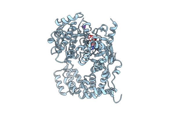 |
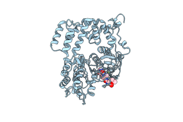
Human G Protein-Coupled Receptor Kinase 5-D311N In Complex With Sangivamycin Soaked In Ph 6
Organism: Homo sapiens
Method: X-RAY DIFFRACTION Release Date: 2025-11-26 Classification: SIGNALING PROTEIN Ligands: SGV, K |
 |
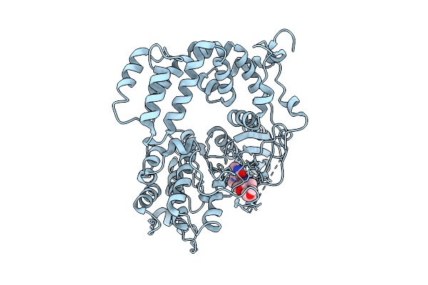
Organism: Homo sapiens
Method: X-RAY DIFFRACTION Release Date: 2025-11-19 Classification: TRANSFERASE Ligands: GSH, FBP |
 |
Structural Basis For The Recognition Of Blood Group Trisaccharides By Tulane Virus
Organism: Rhesus macaque recovirus
Method: ELECTRON MICROSCOPY Release Date: 2025-11-19 Classification: VIRAL PROTEIN |
 |
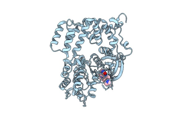
Crystal Structure Of Human G Protein-Coupled Receptor Kinase 5 In Complex With Grl098-22
Organism: Homo sapiens
Method: X-RAY DIFFRACTION Release Date: 2025-11-12 Classification: SIGNALING PROTEIN/INHIBITOR Ligands: A1ARA |
 |
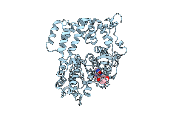
Crystal Structure Of Human G Protein-Coupled Receptor Kinase 5 In Complex With Grl080-22
Organism: Homo sapiens
Method: X-RAY DIFFRACTION Release Date: 2025-11-12 Classification: SIGNALING PROTEIN/INHIBITOR Ligands: A1ARB |
 |
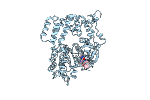
Crystal Structure Of Human G Protein-Coupled Receptor Kinase 5 In Complex With Grl077-22
Organism: Homo sapiens
Method: X-RAY DIFFRACTION Release Date: 2025-11-12 Classification: SIGNALING PROTEIN/INHIBITOR Ligands: A1ARC |
 |
Crystal Structure Of Human G Protein-Coupled Receptor Kinase 5 In Complex With Grl050-22
Organism: Homo sapiens
Method: X-RAY DIFFRACTION Release Date: 2025-11-12 Classification: SIGNALING PROTEIN/INHIBITOR Ligands: A1ARD |
 |
Crystal Structure Of Human G Protein-Coupled Receptor Kinase 5 In Complex With Grl030-22
Organism: Homo sapiens
Method: X-RAY DIFFRACTION Release Date: 2025-11-12 Classification: SIGNALING PROTEIN/INHIBITOR Ligands: A1ARE |