
Search Count: 21
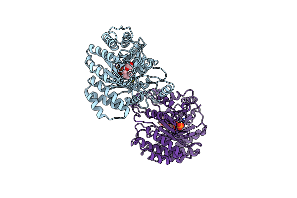 |
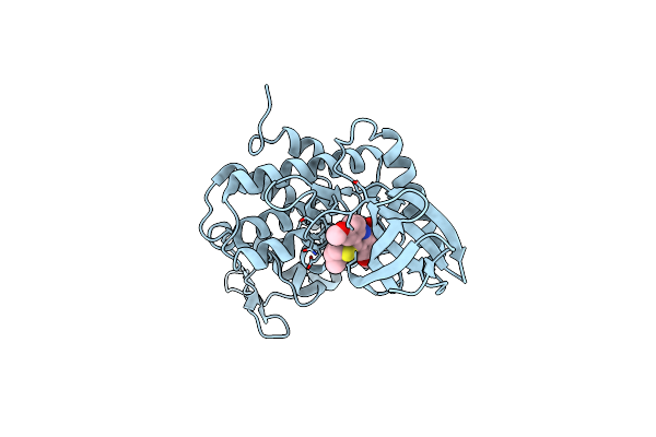
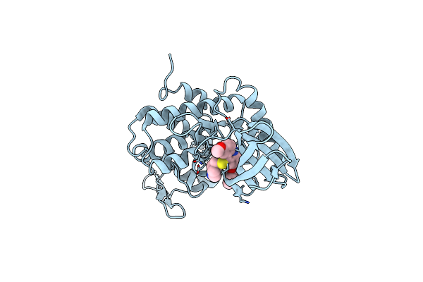
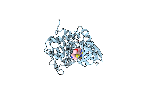
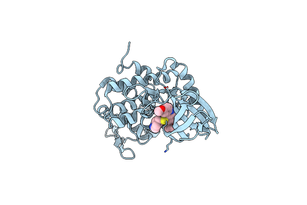
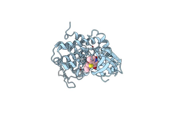
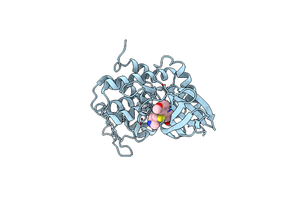
Organism: Fusarium tricinctum
Method: X-RAY DIFFRACTION Resolution:2.27 Å Release Date: 2024-12-11 Classification: TRANSFERASE Ligands: PPV, TOE, MOE |
 |
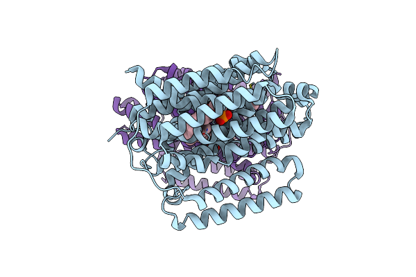
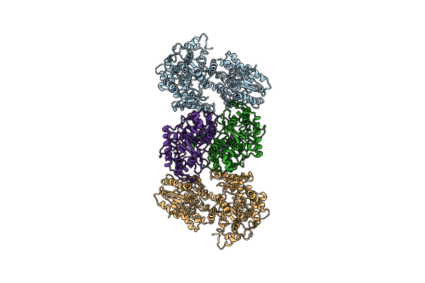
Organism: Mycobacterium tuberculosis h37rv
Method: ELECTRON MICROSCOPY Release Date: 2024-12-04 Classification: PROTEIN BINDING Ligands: A1AQR |
 |
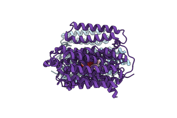
Structure Of The Multidrug Efflux Pump Efpa From M. Tuberculosis Complexed With Lipids
Organism: Mycobacterium tuberculosis
Method: ELECTRON MICROSCOPY Release Date: 2024-11-27 Classification: MEMBRANE PROTEIN Ligands: Y86, Y8F |
 |
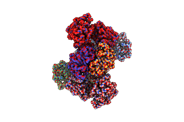
Organism: Planctomycetes bacterium
Method: ELECTRON MICROSCOPY Release Date: 2022-03-16 Classification: RNA BINDING PROTEIN/RNA/DNA |
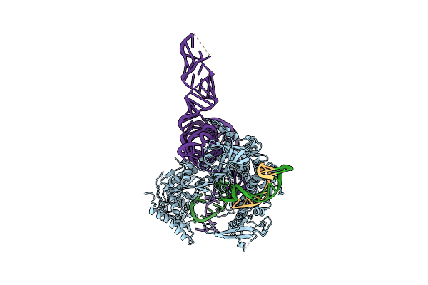 |
Organism: Planctomycetes bacterium
Method: ELECTRON MICROSCOPY Release Date: 2022-03-16 Classification: RNA BINDING PROTEIN-DNA-RNA |
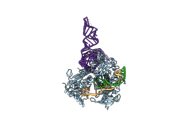 |
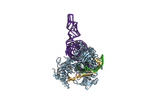
Plmcasx-Sgrnav1-Dsdna Ternary Complex At Nts Loading State With Flexible H2 Domain
Organism: Planctomycetes bacterium
Method: ELECTRON MICROSCOPY Release Date: 2022-03-16 Classification: RNA BINDING PROTEIN/RNA/DNA |
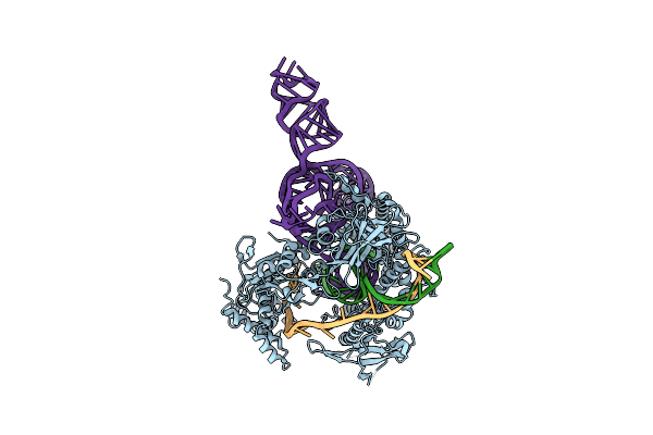 |
Organism: Planctomycetes bacterium
Method: ELECTRON MICROSCOPY Release Date: 2022-03-16 Classification: RNA BINDING PROTEIN/RNA/DNA |
 |
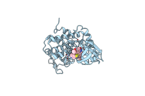
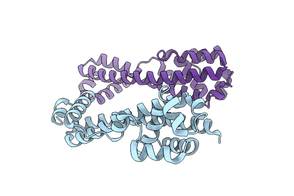
Organism: Homo sapiens
Method: X-RAY DIFFRACTION Resolution:2.65 Å Release Date: 2021-12-01 Classification: CELL CYCLE Ligands: H96 |
 |
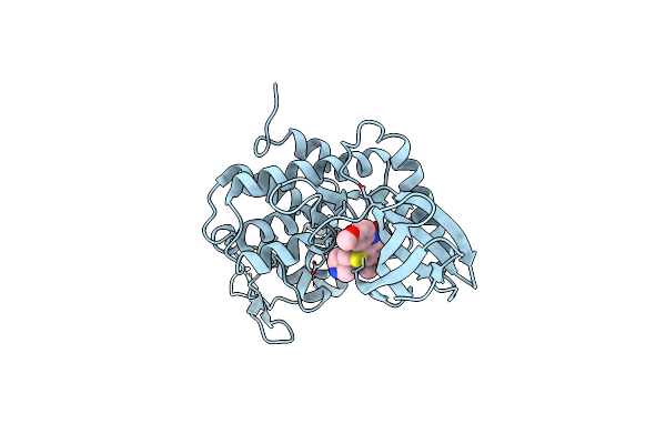
Organism: Homo sapiens
Method: X-RAY DIFFRACTION Resolution:2.50 Å Release Date: 2021-11-24 Classification: CELL CYCLE Ligands: H80 |
 |
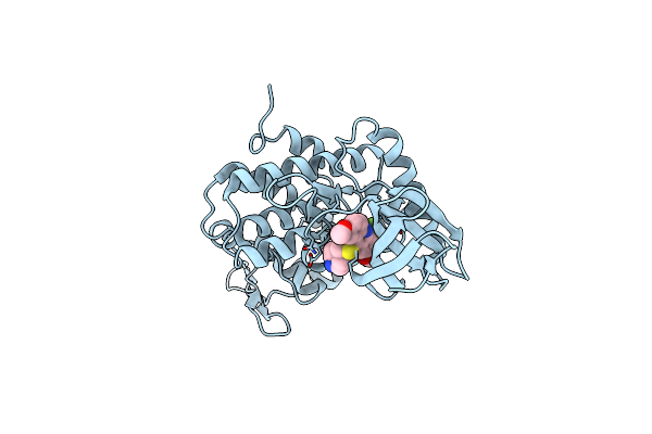
Organism: Homo sapiens
Method: X-RAY DIFFRACTION Resolution:2.19 Å Release Date: 2021-11-17 Classification: CELL CYCLE Ligands: H7F |
 |
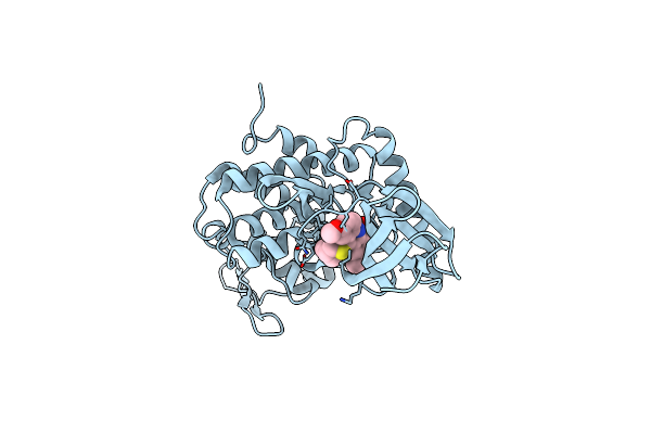
Organism: Homo sapiens
Method: X-RAY DIFFRACTION Resolution:2.59 Å Release Date: 2021-11-17 Classification: CELL CYCLE Ligands: H99 |
 |
Organism: Homo sapiens
Method: X-RAY DIFFRACTION Resolution:2.49 Å Release Date: 2021-11-17 Classification: CELL CYCLE Ligands: H7L |
 |
Organism: Homo sapiens
Method: X-RAY DIFFRACTION Resolution:2.34 Å Release Date: 2021-11-17 Classification: CELL CYCLE Ligands: H7O |
 |
Organism: Homo sapiens
Method: X-RAY DIFFRACTION Resolution:2.38 Å Release Date: 2021-11-17 Classification: CELL CYCLE Ligands: H7R |
 |
Organism: Homo sapiens
Method: X-RAY DIFFRACTION Resolution:3.29 Å Release Date: 2021-11-17 Classification: CELL CYCLE Ligands: H7U |
 |
Organism: Homo sapiens
Method: X-RAY DIFFRACTION Resolution:2.68 Å Release Date: 2021-11-17 Classification: CELL CYCLE Ligands: H7X |
 |
Organism: Homo sapiens
Method: X-RAY DIFFRACTION Resolution:2.58 Å Release Date: 2021-11-10 Classification: CELL CYCLE Ligands: H5R |
 |
Organism: Homo sapiens
Method: ELECTRON MICROSCOPY Release Date: 2018-06-20 Classification: LYASE,HYDROLASE/TRANSFERASE |
 |
Organism: Homo sapiens
Method: ELECTRON MICROSCOPY Release Date: 2018-06-20 Classification: Liase, Oxidoreductase/Transferase |
 |
Organism: Saccharomyces cerevisiae
Method: X-RAY DIFFRACTION Resolution:1.70 Å Release Date: 2009-09-08 Classification: PROTEIN TRANSPORT, ENDOCYTOSIS |