Planned Maintenance: Some services may turn out to be unavailable from 15th January, 2026 to 16th January, 2026. We apologize for the inconvenience!

Planned Maintenance: Some services may turn out to be unavailable from 15th January, 2026 to 16th January, 2026. We apologize for the inconvenience!
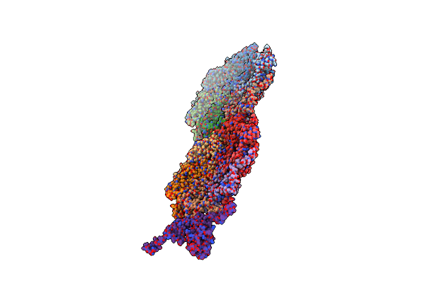 |
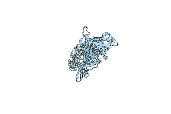
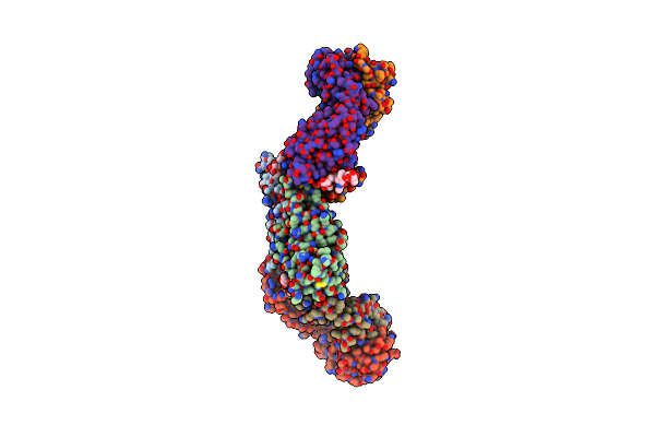
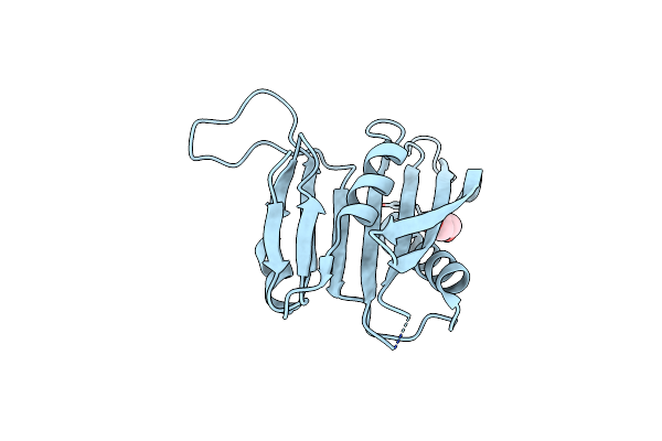
Organism: Escherichia phage dt57c
Method: ELECTRON MICROSCOPY Release Date: 2023-12-13 Classification: VIRUS |
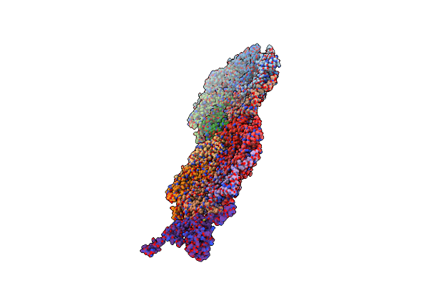 |
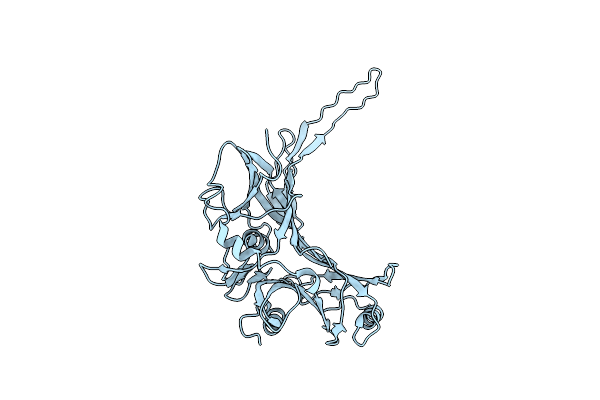
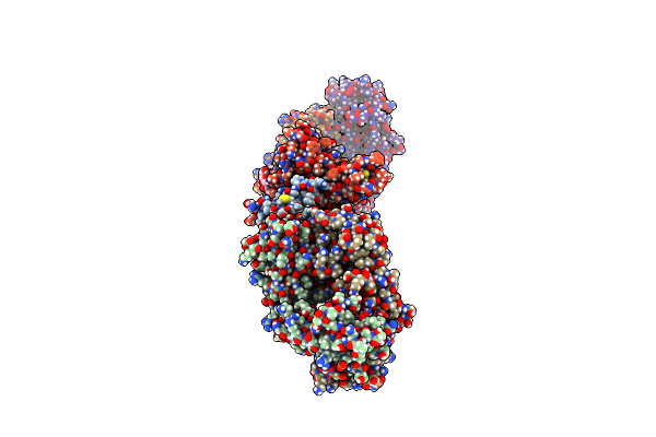
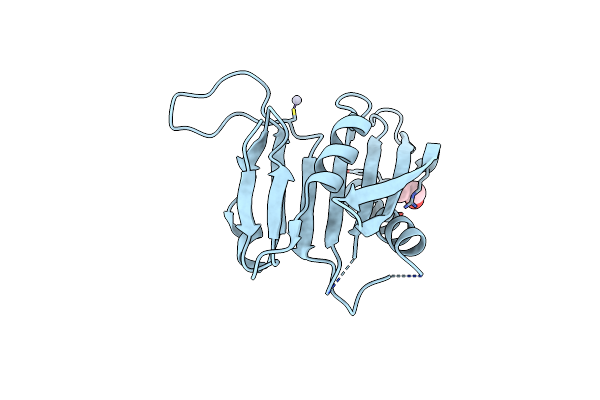
Organism: Escherichia phage dt57c
Method: ELECTRON MICROSCOPY Release Date: 2023-12-13 Classification: VIRUS |
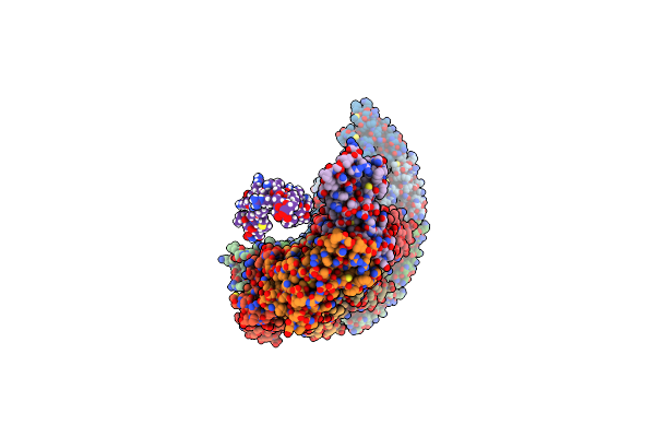 |
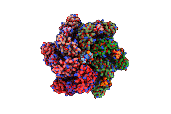
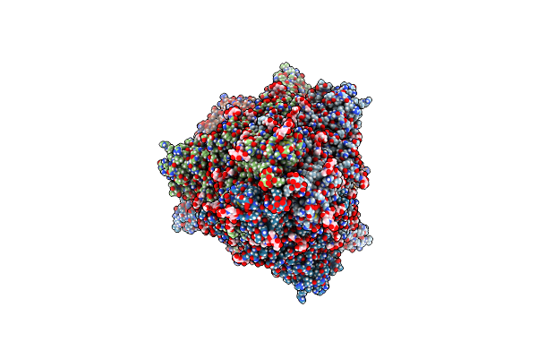
Organism: Escherichia phage dt57c
Method: ELECTRON MICROSCOPY Release Date: 2023-12-13 Classification: VIRAL PROTEIN |
 |
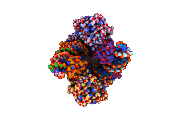
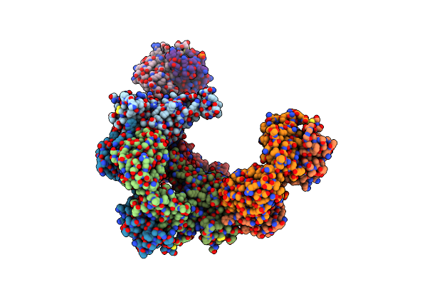
Organism: Escherichia phage dt57c
Method: ELECTRON MICROSCOPY Release Date: 2023-12-13 Classification: VIRAL PROTEIN |
 |
Organism: Escherichia phage dt57c
Method: ELECTRON MICROSCOPY Release Date: 2023-12-13 Classification: VIRAL PROTEIN |
 |
Organism: Escherichia phage dt57c
Method: ELECTRON MICROSCOPY Release Date: 2023-12-13 Classification: VIRAL PROTEIN |
 |
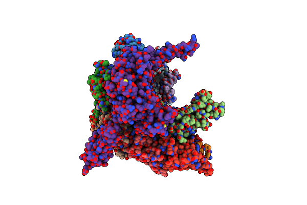
Organism: [scytonema hofmanni] utex 2349, Synthetic construct
Method: ELECTRON MICROSCOPY Release Date: 2022-08-10 Classification: DNA BINDING PROTEIN/DNA Ligands: ANP, MG |
 |
Organism: [scytonema hofmanni] utex 2349, Synthetic construct
Method: ELECTRON MICROSCOPY Release Date: 2022-08-10 Classification: DNA BINDING PROTEIN/DNA Ligands: MG |
 |
Organism: Severe acute respiratory syndrome coronavirus 2, Homo sapiens
Method: ELECTRON MICROSCOPY Release Date: 2022-03-23 Classification: VIRAL PROTEIN/IMMUNE SYSTEM Ligands: NAG |
 |
Crystal Structure Of Sars-Cov-2 Receptor Binding Domain In Complex With The Monoclonal Antibody M31A7
Organism: Severe acute respiratory syndrome coronavirus 2, Mus musculus
Method: X-RAY DIFFRACTION Resolution:3.20 Å Release Date: 2022-03-16 Classification: VIRAL PROTEIN/IMMUNE SYSTEM |
 |
Organism: Severe acute respiratory syndrome coronavirus 2
Method: ELECTRON MICROSCOPY Release Date: 2020-08-26 Classification: VIRAL PROTEIN Ligands: NAG |
 |
Association Of Three Complexes Of Largely Structurally Disordered Spike Ectodomain With Bound Ey6A Fab
Organism: Severe acute respiratory syndrome coronavirus 2, Homo sapiens
Method: ELECTRON MICROSCOPY Release Date: 2020-07-29 Classification: VIRAL PROTEIN Ligands: NAG |
 |
Association Of Two Complexes Of Largely Structurally Disordered Spike Ectodomain With Bound Ey6A Fab
Organism: Severe acute respiratory syndrome coronavirus 2, Homo sapiens
Method: ELECTRON MICROSCOPY Release Date: 2020-07-08 Classification: IMMUNE SYSTEM Ligands: NAG |
 |
Sars-Cov-2 Spike Glycoprotein In Complex With A Neutralizing Antibody Ey6A Fab
Organism: Severe acute respiratory syndrome coronavirus 2, Homo sapiens
Method: ELECTRON MICROSCOPY Release Date: 2020-07-01 Classification: VIRAL PROTEIN Ligands: NAG |
 |
Crystal Structure Of Receptor Binding Domain Of Sars-Cov-2 Spike Glycoprotein In Ternary Complex With Ey6A Fab And A Nanobody.
Organism: Severe acute respiratory syndrome coronavirus 2, Lama glama, Homo sapiens
Method: X-RAY DIFFRACTION Resolution:2.65 Å Release Date: 2020-06-24 Classification: VIRAL PROTEIN Ligands: NAG, CL, MG |
 |
Crystal Structure Of Receptor Binding Domain Of Sars-Cov-2 Spike Glycoprotein In Complex With Ey6A Fab
Organism: Severe acute respiratory syndrome coronavirus 2, Homo sapiens
Method: X-RAY DIFFRACTION Resolution:3.80 Å Release Date: 2020-06-24 Classification: VIRAL PROTEIN/IMMUNE SYSTEM Ligands: NAG, PO4 |
 |
Crystal Structure Of Apo Form Fibronectin-Binding Protein Apa From Mycobacterium Tuberculosis
Organism: Mycobacterium tuberculosis (strain atcc 25618 / h37rv)
Method: X-RAY DIFFRACTION Resolution:1.55 Å Release Date: 2019-05-29 Classification: PROTEIN BINDING Ligands: GOL |
 |
Crystal Structure Of Fibronectin-Binding Protein Apa Mutant From Mycobacterium Tuberculosis
Organism: Mycobacterium tuberculosis (strain atcc 25618 / h37rv)
Method: X-RAY DIFFRACTION Resolution:1.77 Å Release Date: 2019-05-29 Classification: PROTEIN BINDING Ligands: HG, GOL |