
Search Count: 363
 |
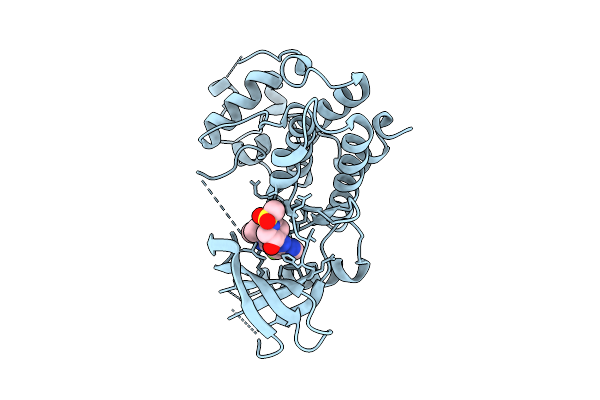
Organism: Homo sapiens
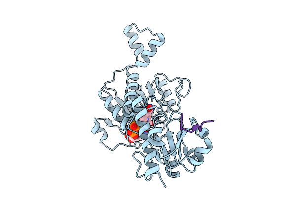
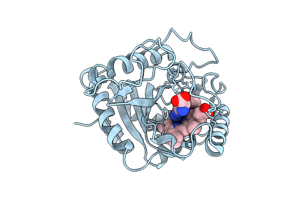
Method: X-RAY DIFFRACTION Resolution:2.20 Å Release Date: 2026-01-28 Classification: PROTEIN BINDING Ligands: NAD |
 |
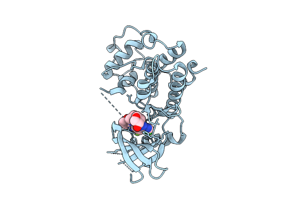
Organism: Homo sapiens
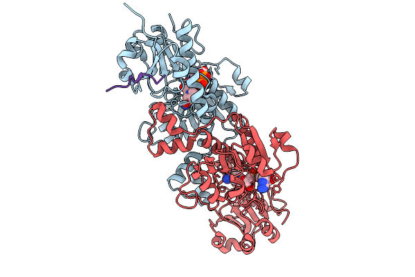
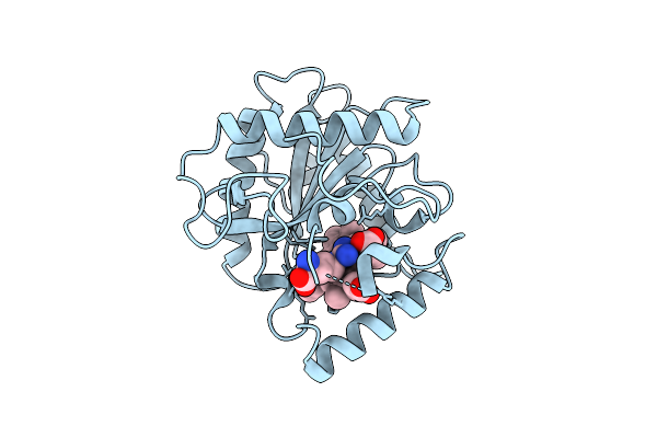
Method: X-RAY DIFFRACTION Resolution:1.85 Å Release Date: 2026-01-21 Classification: PROTEIN BINDING Ligands: NAD |
 |
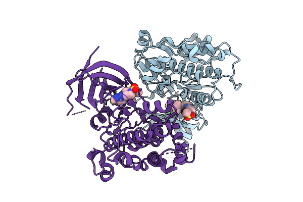
Organism: Homo sapiens, Mus musculus
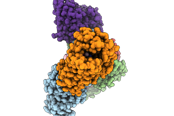
Method: ELECTRON MICROSCOPY Resolution:2.84 Å Release Date: 2026-01-14 Classification: MEMBRANE PROTEIN Ligands: CLR, A1EK5 |
 |
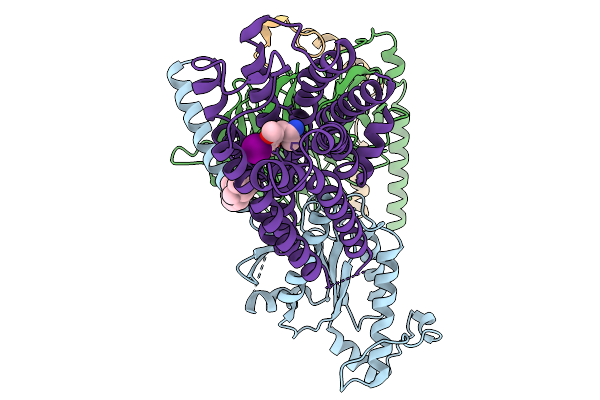
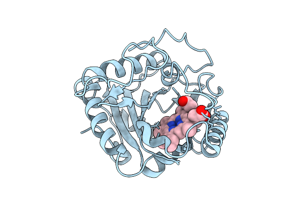
Organism: Homo sapiens
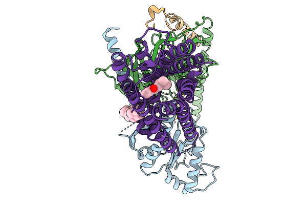
Method: ELECTRON MICROSCOPY Resolution:2.62 Å Release Date: 2026-01-14 Classification: MEMBRANE PROTEIN Ligands: 91Q, CLR |
 |
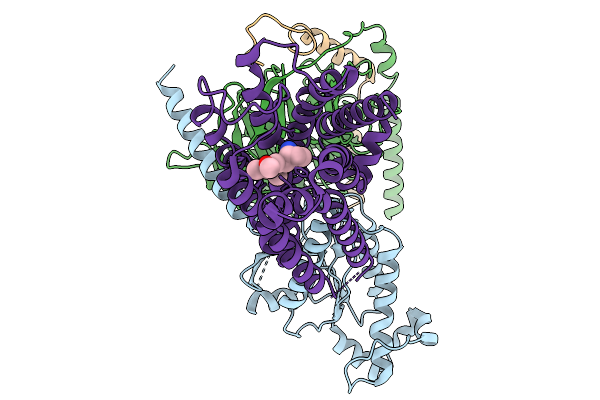
Organism: Homo sapiens
Method: ELECTRON MICROSCOPY Resolution:2.76 Å Release Date: 2026-01-14 Classification: MEMBRANE PROTEIN Ligands: A1EK5, CLR |
 |
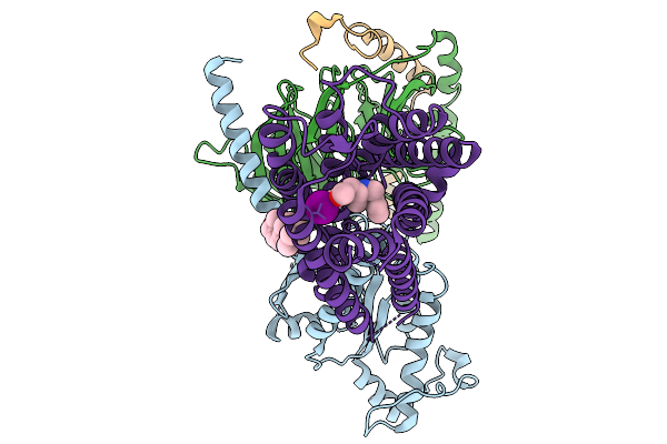
Organism: Homo sapiens
Method: ELECTRON MICROSCOPY Resolution:3.27 Å Release Date: 2026-01-14 Classification: MEMBRANE PROTEIN Ligands: A1EK8 |
 |
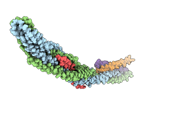
Organism: Homo sapiens
Method: ELECTRON MICROSCOPY Resolution:2.98 Å Release Date: 2026-01-14 Classification: MEMBRANE PROTEIN Ligands: CLR, A1ELA |
 |
Organism: Saccharomyces cerevisiae s288c
Method: X-RAY DIFFRACTION Resolution:3.50 Å Release Date: 2026-01-14 Classification: GENE REGULATION |
 |
Organism: Severe acute respiratory syndrome coronavirus 2, Homo sapiens
Method: X-RAY DIFFRACTION Resolution:2.39 Å Release Date: 2026-01-07 Classification: VIRAL PROTEIN/IMMUNE SYSTEM Ligands: NAG |
 |
Organism: Homo sapiens
Method: X-RAY DIFFRACTION Resolution:2.05 Å Release Date: 2025-12-17 Classification: TRANSFERASE/INHIBITOR Ligands: A1CIF |
 |
Organism: Homo sapiens
Method: X-RAY DIFFRACTION Resolution:1.80 Å Release Date: 2025-12-17 Classification: TRANSFERASE/INHIBITOR Ligands: A1AZ4 |
 |
Organism: Homo sapiens
Method: X-RAY DIFFRACTION Resolution:2.11 Å Release Date: 2025-12-17 Classification: TRANSFERASE/INHIBITOR Ligands: A1CIF |
 |
Organism: Streptomyces monomycini
Method: X-RAY DIFFRACTION Resolution:2.06 Å Release Date: 2025-12-10 Classification: BIOSYNTHETIC PROTEIN Ligands: HEM |
 |
Organism: Streptomyces monomycini
Method: X-RAY DIFFRACTION Resolution:1.83 Å Release Date: 2025-12-10 Classification: BIOSYNTHETIC PROTEIN Ligands: HEM, ARG |
 |
Organism: Streptomyces monomycini
Method: X-RAY DIFFRACTION Resolution:1.47 Å Release Date: 2025-12-10 Classification: BIOSYNTHETIC PROTEIN Ligands: HEM, ARG |
 |
Organism: Streptomyces monomycini
Method: X-RAY DIFFRACTION Resolution:1.41 Å Release Date: 2025-12-10 Classification: BIOSYNTHETIC PROTEIN Ligands: HEM, ARG |
 |
Organism: Streptomyces monomycini
Method: X-RAY DIFFRACTION Resolution:1.49 Å Release Date: 2025-12-10 Classification: BIOSYNTHETIC PROTEIN Ligands: ARG, HEM |
 |
Organism: Streptomyces monomycini
Method: X-RAY DIFFRACTION Resolution:1.60 Å Release Date: 2025-12-10 Classification: BIOSYNTHETIC PROTEIN Ligands: HEM, ARG, PGL |
 |
Crystal Structure Of A Allulose Transcriptional Regulator From Agrobacterium Fabrum
Organism: Agrobacterium fabrum str. c58
Method: X-RAY DIFFRACTION Resolution:2.53 Å Release Date: 2025-11-26 Classification: TRANSCRIPTION Ligands: A1EGE |
 |
Organism: Homo sapiens, Mus musculus, Escherichia coli
Method: ELECTRON MICROSCOPY Resolution:3.13 Å Release Date: 2025-11-26 Classification: MEMBRANE PROTEIN Ligands: 8K3 |