
Search Count: 12
 |
Organism: Escherichia coli (strain k12), Synthetic construct
Method: X-RAY DIFFRACTION Resolution:1.97 Å Release Date: 2019-03-20 Classification: OXIDOREDUCTASE Ligands: CA |
 |
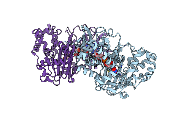
Organism: Escherichia coli (strain k12), Synthetic construct
Method: X-RAY DIFFRACTION Resolution:1.80 Å Release Date: 2019-03-20 Classification: OXIDOREDUCTASE |
 |
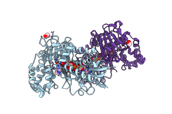
Organism: Escherichia coli (strain k12), Synthetic construct, Homo sapiens
Method: X-RAY DIFFRACTION Resolution:3.30 Å Release Date: 2019-03-20 Classification: OXIDOREDUCTASE |
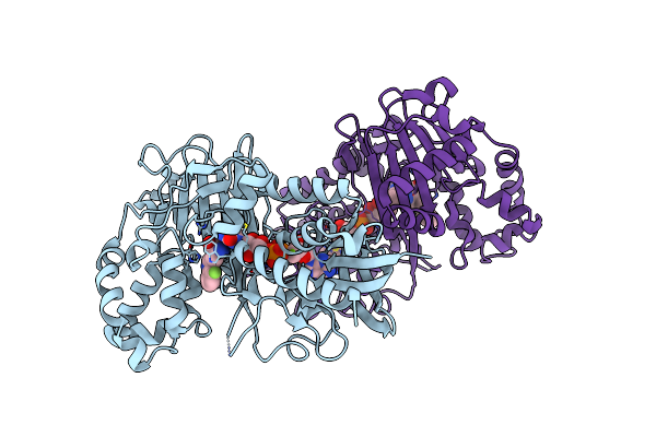 |
Crystal Structure Of L-Tryptophan Oxidase Vioa From Chromobacterium Violaceum In Complex With 4-Fluoro-L-Tryptophan
Organism: Chromobacterium violaceum atcc 12472
Method: X-RAY DIFFRACTION Resolution:3.00 Å Release Date: 2019-02-13 Classification: BIOSYNTHETIC PROTEIN Ligands: FAD, 4FW, MG |
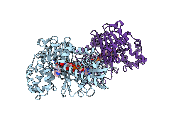 |
Crystal Structure Of L-Tryptophan Oxidase Vioa From Chromobacterium Violaceum In Complex With 5-Methyl-L-Tryptophan
Organism: Chromobacterium violaceum atcc 12472
Method: X-RAY DIFFRACTION Resolution:2.40 Å Release Date: 2019-02-13 Classification: BIOSYNTHETIC PROTEIN Ligands: FAD, D0Q, MG |
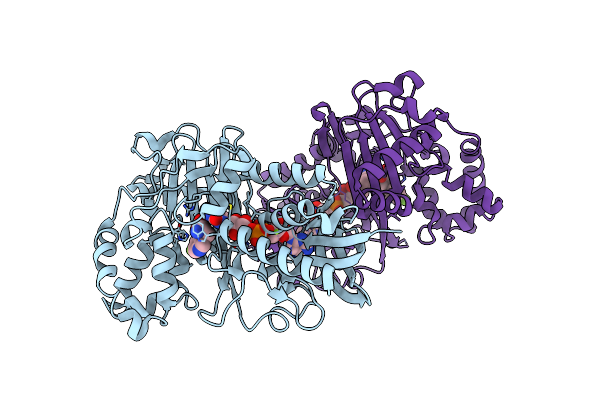 |
Crystal Structure Of L-Tryptophan Oxidase Vioa From Chromobacterium Violaceum In Complex With 6-Fluoro-L-Tryptophan
Organism: Chromobacterium violaceum atcc 12472
Method: X-RAY DIFFRACTION Resolution:2.74 Å Release Date: 2019-02-13 Classification: BIOSYNTHETIC PROTEIN Ligands: FAD, FT6, MG |
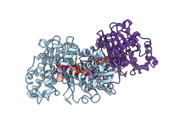 |
Crystal Structure Of L-Tryptophan Oxidase Vioa From Chromobacterium Violaceum In Complex With 7-Methyl-L-Tryptophan
Organism: Chromobacterium violaceum (strain atcc 12472 / dsm 30191 / jcm 1249 / nbrc 12614 / ncimb 9131 / nctc 9757)
Method: X-RAY DIFFRACTION Resolution:2.85 Å Release Date: 2019-02-13 Classification: BIOSYNTHETIC PROTEIN Ligands: FAD, E95, MG |
 |
Crystal Structure Of L-Tryptophan Oxidase Vioa From Chromobacterium Violaceum In Complex With L-Tryptophan
Organism: Chromobacterium violaceum atcc 12472
Method: X-RAY DIFFRACTION Resolution:2.60 Å Release Date: 2018-05-16 Classification: BIOSYNTHETIC PROTEIN Ligands: FAD, TRP, EDO, MG |
 |
Structure-Activity Studies Of Mdm2/Mdm4-Binding Stapled Peptides Comprising Non-Natural Amino Acids
Organism: Homo sapiens, Phage display vector ptdisp
Method: X-RAY DIFFRACTION Resolution:1.66 Å Release Date: 2017-12-27 Classification: ONCOPROTEIN/INHIBITOR |
 |
Organism: Aequorea victoria, Camelus dromedarius, Homo sapiens
Method: X-RAY DIFFRACTION Resolution:3.00 Å Release Date: 2017-12-20 Classification: FLUORESCENT PROTEIN/INHIBITOR Ligands: GOL, PO4, PG4 |
 |
Crystal Structure Of L-Tryptophan Oxidase Vioa From Chromobacterium Violaceum
Organism: Chromobacterium violaceum (strain atcc 12472 / dsm 30191 / jcm 1249 / nbrc 12614 / ncimb 9131 / nctc 9757)
Method: X-RAY DIFFRACTION Resolution:2.60 Å Release Date: 2017-12-06 Classification: BIOSYNTHETIC PROTEIN Ligands: FAD, CL |
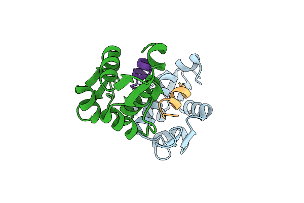 |
Organism: Homo sapiens, Synthetic construct
Method: X-RAY DIFFRACTION Resolution:1.99 Å Release Date: 2014-05-28 Classification: CELL CYCLE |