
Search Count: 13
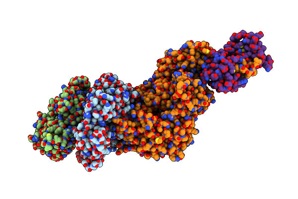 |
Organism: Mus musculus, Oryctolagus cuniculus
Method: ELECTRON MICROSCOPY Release Date: 2025-02-12 Classification: MOTOR PROTEIN Ligands: ADP, MG |
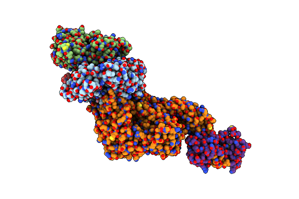 |
Organism: Mus musculus, Oryctolagus cuniculus
Method: ELECTRON MICROSCOPY Release Date: 2025-02-12 Classification: MOTOR PROTEIN Ligands: ADP, MG |
 |
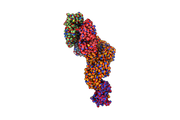
Organism: Mus musculus, Oryctolagus cuniculus
Method: ELECTRON MICROSCOPY Release Date: 2025-02-12 Classification: MOTOR PROTEIN Ligands: ADP, MG |
 |
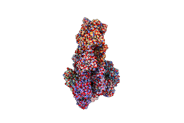
Organism: Mus musculus, Oryctolagus cuniculus
Method: ELECTRON MICROSCOPY Release Date: 2025-02-12 Classification: MOTOR PROTEIN Ligands: ADP, MG |
 |
Organism: Schizosaccharomyces pombe, Oryctolagus cuniculus
Method: ELECTRON MICROSCOPY Release Date: 2024-01-31 Classification: CYTOSOLIC PROTEIN Ligands: ADP, MG, ATP |
 |
Organism: Schizosaccharomyces pombe, Oryctolagus cuniculus
Method: ELECTRON MICROSCOPY Release Date: 2024-01-31 Classification: CYTOSOLIC PROTEIN Ligands: ADP, MG, BEF, ATP |
 |
Organism: Oryctolagus cuniculus
Method: ELECTRON MICROSCOPY Release Date: 2024-01-31 Classification: CYTOSOLIC PROTEIN Ligands: ADP, MG |
 |
Organism: Oryctolagus cuniculus
Method: ELECTRON MICROSCOPY Release Date: 2024-01-31 Classification: CYTOSOLIC PROTEIN Ligands: ADP, MG, BEF |
 |
Organism: Homo sapiens
Method: X-RAY DIFFRACTION Resolution:2.20 Å Release Date: 2021-03-17 Classification: RNA BINDING PROTEIN |
 |
Co-Crystal Structure Of Hiv-1 Tar Rna In Complex With Lab-Evolved Rrm Tbp6.9
Organism: Homo sapiens, Human immunodeficiency virus 1
Method: X-RAY DIFFRACTION Resolution:3.10 Å Release Date: 2020-10-14 Classification: RNA BINDING PROTEIN/RNA Ligands: MG |
 |
Co-Crystal Structure Of Hiv-1 Tar Rna In Complex With Lab-Evolved Rrm Tbp6.7 Mutant
Organism: Homo sapiens, Human immunodeficiency virus 1
Method: X-RAY DIFFRACTION Resolution:2.60 Å Release Date: 2020-10-14 Classification: RNA BINDING PROTEIN/RNA |
 |
Organism: Homo sapiens, Human immunodeficiency virus 1
Method: X-RAY DIFFRACTION Resolution:1.71 Å Release Date: 2020-10-14 Classification: RNA BINDING PROTEIN/RNA |
 |
Co-Crystal Structure Of Hiv-1 Tar Rna In Complex With Lab-Evolved Rrm Tbp6.3
Organism: Homo sapiens, Human immunodeficiency virus 1
Method: X-RAY DIFFRACTION Resolution:2.35 Å Release Date: 2020-10-14 Classification: RNA BINDING PROTEIN/RNA |