
Search Count: 20
 |
Organism: Homo sapiens, Escherichia coli
Method: ELECTRON MICROSCOPY Release Date: 2024-02-21 Classification: MEMBRANE PROTEIN Ligands: CA, NAG, POV, 9Z9, EPJ |
 |
Organism: Homo sapiens, Escherichia coli
Method: ELECTRON MICROSCOPY Release Date: 2024-02-21 Classification: MEMBRANE PROTEIN Ligands: XG3, NAG, EPJ, 9Z9, POV |
 |
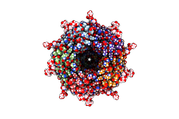
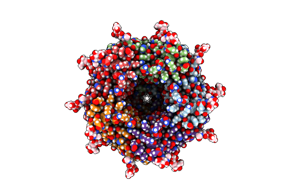
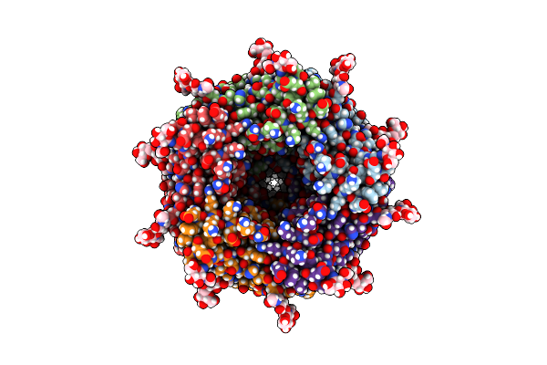
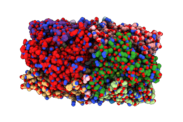
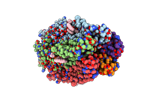
Alpha7-Nicotinic Acetylcholine Receptor Bound To Epibatidine And Ivermectin
Organism: Homo sapiens
Method: ELECTRON MICROSCOPY Release Date: 2024-02-21 Classification: MEMBRANE PROTEIN Ligands: CA, NAG, 9Z9, EPJ, IVM, POV |
 |
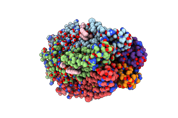
Organism: Homo sapiens
Method: ELECTRON MICROSCOPY Release Date: 2024-02-21 Classification: MEMBRANE PROTEIN Ligands: CA, NAG, POV, 9Z9, EPJ, YLR |
 |
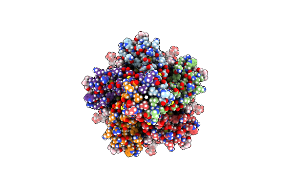
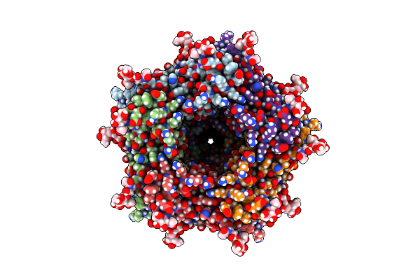
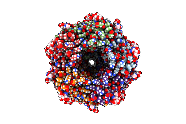
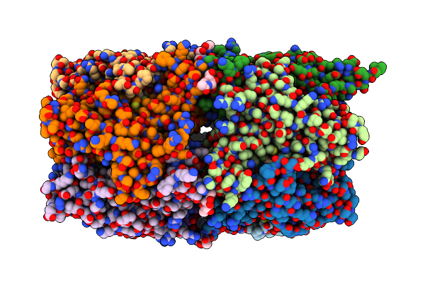
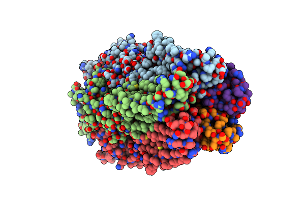
Alpha7-Nicotinic Acetylcholine Receptor Bound To Epibatidine And Pnu-120596
Organism: Homo sapiens
Method: ELECTRON MICROSCOPY Release Date: 2024-02-21 Classification: MEMBRANE PROTEIN Ligands: CA, NAG, POV, 9Z9, EPJ, I34 |
 |
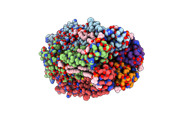
Organism: Homo sapiens
Method: ELECTRON MICROSCOPY Release Date: 2024-02-21 Classification: MEMBRANE PROTEIN Ligands: POV, 9Z9, NAG, YLI |
 |
Organism: Homo sapiens
Method: ELECTRON MICROSCOPY Release Date: 2024-02-21 Classification: MEMBRANE PROTEIN Ligands: CA, NAG, POV, 9Z9, EPJ, YLI |
 |
Organism: Homo sapiens
Method: ELECTRON MICROSCOPY Release Date: 2024-02-21 Classification: MEMBRANE PROTEIN Ligands: NAG, R16, 9Z9, POV |
 |
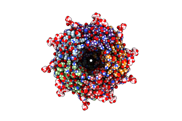
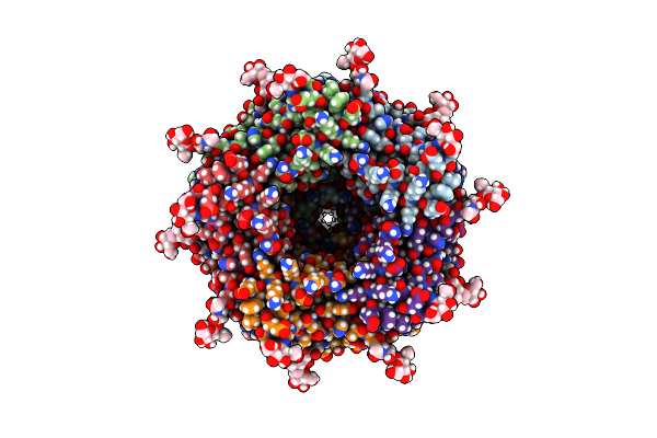
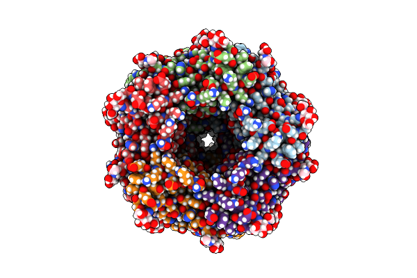
Alpha7-Nicotinic Acetylcholine Receptor Time Resolved Bound To Epibatidine And Pnu-120596 Desensitized Intermediate State
Organism: Homo sapiens
Method: ELECTRON MICROSCOPY Release Date: 2024-02-21 Classification: MEMBRANE PROTEIN Ligands: CA, NAG, POV, 9Z9, EPJ, I34 |
 |
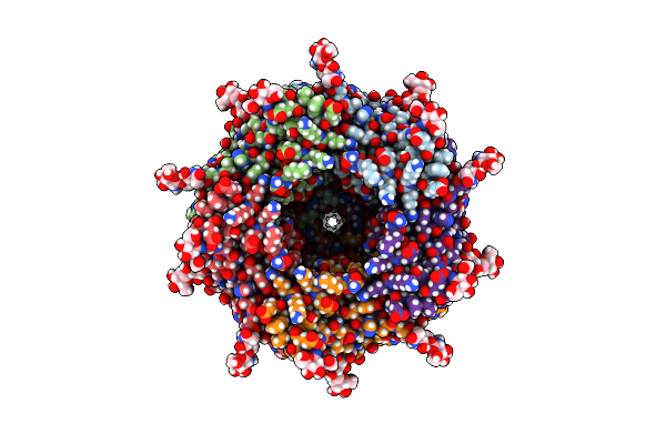
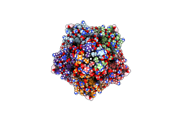
Alpha7-Nicotinic Acetylcholine Receptor Time Resolved Bound To Epibatidine And Pnu-120596 Asymmetric State 1
Organism: Homo sapiens
Method: ELECTRON MICROSCOPY Release Date: 2024-02-21 Classification: MEMBRANE PROTEIN Ligands: NAG, EPJ, I34, CA |
 |
Alpha7-Nicotinic Acetylcholine Receptor Time Resolved Bound To Epibatidine And Pnu-120596 Asymmetric State 2
Organism: Homo sapiens
Method: ELECTRON MICROSCOPY Release Date: 2024-02-21 Classification: MEMBRANE PROTEIN Ligands: NAG, EPJ, I34, CA |
 |
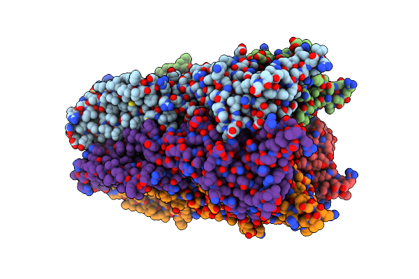
X-Ray Structure Of Acetylcholine Binding Protein (Achbp) In Complex With 6-(4-Methoxyphenyl)-N4-Octylpyrimidine-2,4-Diamine
Organism: Lymnaea stagnalis
Method: X-RAY DIFFRACTION Resolution:2.70 Å Release Date: 2014-07-16 Classification: Acetylcholine-Binding Protein Ligands: KK1, NAG, PO4 |
 |
X-Ray Structure Of Acetylcholine Binding Protein (Achbp) In Complex With 4-(Morpholin-4-Yl)-6-[4-(Trifluoromethyl)Phenyl]Pyrimidin-2-Amine
Organism: Lymnaea stagnalis
Method: X-RAY DIFFRACTION Resolution:2.98 Å Release Date: 2014-07-16 Classification: Acetylcholine-Binding Protein Ligands: KK2, NAG, PO4 |
 |
X-Ray Structure Of Acetylcholine Binding Protein (Achbp) In Complex With 4-(4-Methylpiperidin-1-Yl)-6-(4-(Trifluoromethyl)Phenyl)Pyrimidin-2-Amine
Organism: Lymnaea stagnalis
Method: X-RAY DIFFRACTION Resolution:2.10 Å Release Date: 2014-07-16 Classification: Acetylcholine-Binding Protein Ligands: PO4, NAG, KK3 |
 |
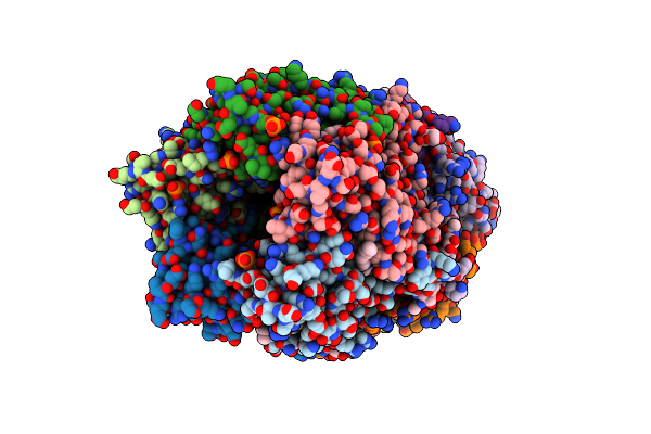
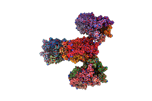
The Glic-His10 Wild-Type Structure In Equilibrium Between The Open And Locally-Closed (Lc) Forms
Organism: Gloeobacter violaceus
Method: X-RAY DIFFRACTION Resolution:3.35 Å Release Date: 2013-12-25 Classification: TRANSPORT PROTEIN Ligands: NI |
 |
Organism: Gloeobacter violaceus
Method: X-RAY DIFFRACTION Resolution:4.35 Å Release Date: 2013-12-25 Classification: TRANSPORT PROTEIN |
 |
Structure Of Desflurane Bound To A Pentameric Ligand-Gated Ion Channel, Glic
Organism: Gloeobacter violaceus
Method: X-RAY DIFFRACTION Resolution:3.20 Å Release Date: 2011-01-19 Classification: MEMBRANE PROTEIN, TRANSPORT PROTEIN Ligands: LMT, DSF, PLC |
 |
Organism: Gloeobacter violaceus
Method: X-RAY DIFFRACTION Resolution:3.30 Å Release Date: 2011-01-19 Classification: MEMBRANE PROTEIN, TRANSPORT PROTEIN Ligands: LMT, PFL, PLC |
 |
Structure Of The A237F Mutant Of The Pentameric Ligand Gated Ion Channel From Gloeobacter Violaceus
Organism: Gloeobacter violaceus
Method: X-RAY DIFFRACTION Resolution:3.15 Å Release Date: 2010-04-28 Classification: MEMBRANE PROTEIN, TRANSPORT PROTEIN |
 |
Organism: Gloeobacter violaceus
Method: X-RAY DIFFRACTION Resolution:2.90 Å Release Date: 2008-11-04 Classification: MEMBRANE PROTEIN, TRANSPORT PROTEIN Ligands: LMT, PC1 |