
Search Count: 344
 |
Organism: Homo sapiens
Method: X-RAY DIFFRACTION Release Date: 2025-11-12 Classification: HYDROLASE Ligands: ZN, CA, NAG, A1JCF, PO4 |
 |
Organism: Homo sapiens
Method: X-RAY DIFFRACTION Release Date: 2025-11-12 Classification: HYDROLASE Ligands: ZN, CA, NAG, A1JCG, PO4 |
 |
Organism: Homo sapiens
Method: X-RAY DIFFRACTION Release Date: 2025-11-12 Classification: HYDROLASE Ligands: ZN, CA, A1JCH, PO4 |
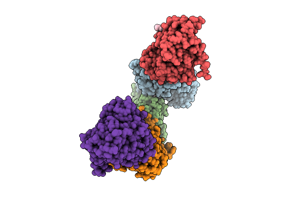 |
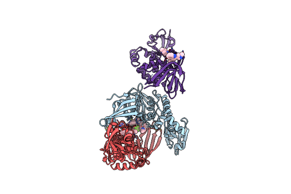
Crystal Structure Of Egfr Exon20 Insertion Mutant In Complex With Enozertinib (Oric-114)
Organism: Homo sapiens
Method: X-RAY DIFFRACTION Release Date: 2025-11-12 Classification: SIGNALING PROTEIN Ligands: A1L8T |
 |
Organism: Homo sapiens
Method: X-RAY DIFFRACTION Release Date: 2025-07-30 Classification: HYDROLASE Ligands: A1CE0, CA |
 |
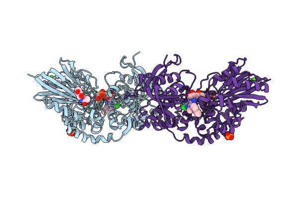
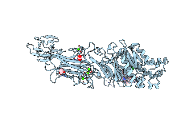
Potent Inhibition Of The Protein Arginine Deiminases (Pad1-4) By Targeting A Ca2+ Dependent Allosteric Binding Site
Organism: Homo sapiens
Method: X-RAY DIFFRACTION Resolution:1.77 Å Release Date: 2025-05-28 Classification: HYDROLASE Ligands: CA, A1A8O |
 |
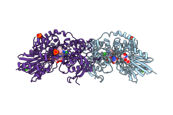
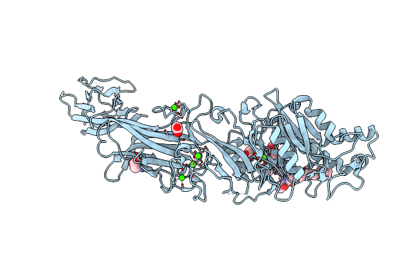
Inhibiting Peptidylarginine Deiminases (Pad1-4) By Targeting A Ca2+ Dependent Allosteric Binding Site
Organism: Homo sapiens
Method: X-RAY DIFFRACTION Resolution:2.44 Å Release Date: 2025-05-28 Classification: HYDROLASE/HYDROLASE INHIBITOR Ligands: CA, A1BZK |
 |
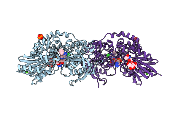
Potent Inhibition Of The Protein Arginine Deiminases (Pad1-4) By Targeting A Ca2+ Dependent Allosteric Binding Site
Organism: Homo sapiens
Method: X-RAY DIFFRACTION Resolution:2.41 Å Release Date: 2025-05-28 Classification: HYDROLASE/HYDROLASE INHIBITOR Ligands: A1A9P, 3YZ |
 |
Organism: Severe acute respiratory syndrome coronavirus 2
Method: ELECTRON MICROSCOPY Resolution:2.90 Å Release Date: 2025-03-19 Classification: VIRAL PROTEIN/RNA Ligands: ZN, MG |
 |
Organism: Severe acute respiratory syndrome coronavirus 2
Method: ELECTRON MICROSCOPY Release Date: 2024-12-04 Classification: VIRAL PROTEIN Ligands: NAG |
 |
Organism: Homo sapiens
Method: X-RAY DIFFRACTION Resolution:1.64 Å Release Date: 2024-10-09 Classification: HYDROLASE Ligands: A1AJA, ACT, CA |
 |
Organism: Homo sapiens
Method: X-RAY DIFFRACTION Resolution:1.95 Å Release Date: 2024-10-09 Classification: HYDROLASE Ligands: MPD, A1AJB, ACT, CA |
 |
Organism: Homo sapiens
Method: X-RAY DIFFRACTION Resolution:2.00 Å Release Date: 2024-10-09 Classification: HYDROLASE Ligands: ACT, CA, A1AJC |
 |
Organism: Severe acute respiratory syndrome coronavirus 2
Method: X-RAY DIFFRACTION Resolution:2.60 Å Release Date: 2024-10-02 Classification: VIRAL PROTEIN Ligands: A1AZ1, TFA, ZN |
 |
Organism: Coxsackievirus b3
Method: X-RAY DIFFRACTION Resolution:2.10 Å Release Date: 2024-08-14 Classification: VIRAL PROTEIN |
 |
Organism: Coxsackievirus b4
Method: X-RAY DIFFRACTION Resolution:2.01 Å Release Date: 2024-08-14 Classification: VIRAL PROTEIN |
 |
Organism: Canis lupus familiaris
Method: X-RAY DIFFRACTION Resolution:1.85 Å Release Date: 2024-05-22 Classification: CYTOKINE |
 |
Organism: Homo sapiens
Method: X-RAY DIFFRACTION Resolution:1.59 Å Release Date: 2024-05-08 Classification: BIOSYNTHETIC PROTEIN Ligands: SO4, GOL |
 |
Organism: Acinetobacter genomosp. 16bj
Method: ELECTRON MICROSCOPY Release Date: 2024-03-06 Classification: CELL ADHESION |
 |
Organism: Bacteria abnormis, Acinetobacter phage ap205
Method: ELECTRON MICROSCOPY Release Date: 2024-03-06 Classification: VIRUS/RNA |