
Search Count: 75
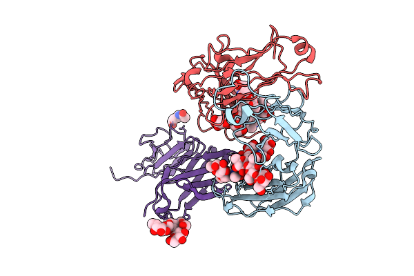 |
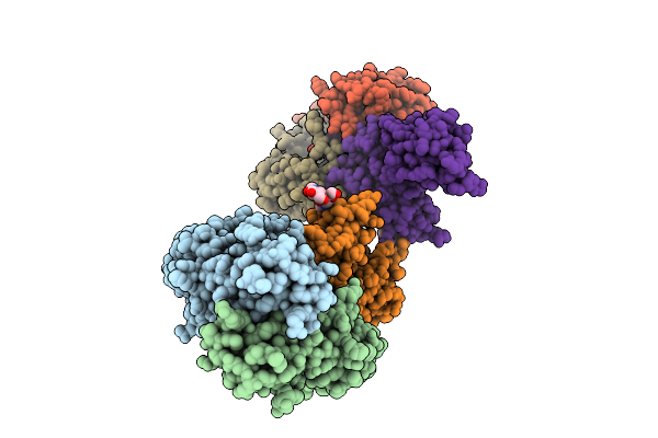
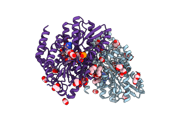
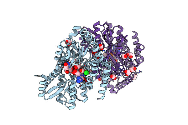
Crystal Structure Of Rhesus Macaque (Macaca Mulatta) Igg1 Fc Fragment- Fc-Gamma Receptor Iia Complex P131 Variant
Organism: Macaca mulatta
Method: X-RAY DIFFRACTION Release Date: 2025-12-03 Classification: IMMUNE SYSTEM Ligands: NAG |
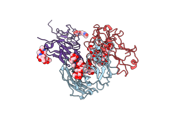 |
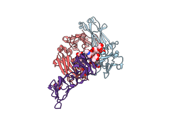
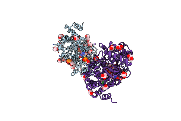
Crystal Structure Of Rhesus Macaque (Macaca Mulatta) Igg1 Fc Fragment- Fc-Gamma Receptor Iia Complex H131 Variant
Organism: Macaca mulatta
Method: X-RAY DIFFRACTION Release Date: 2025-12-03 Classification: IMMUNE SYSTEM |
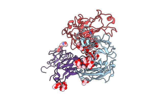 |
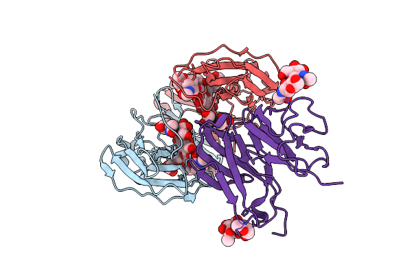
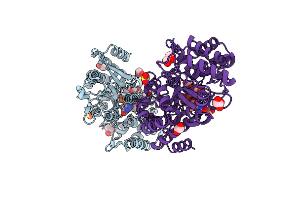
Crystal Structure Of Rhesus Macaque (Macaca Mulatta) Igg2 Fc Fragment- Fc-Gamma Receptor Iia Complex H131 Variant
Organism: Macaca mulatta
Method: X-RAY DIFFRACTION Release Date: 2025-12-03 Classification: IMMUNE SYSTEM Ligands: MPD |
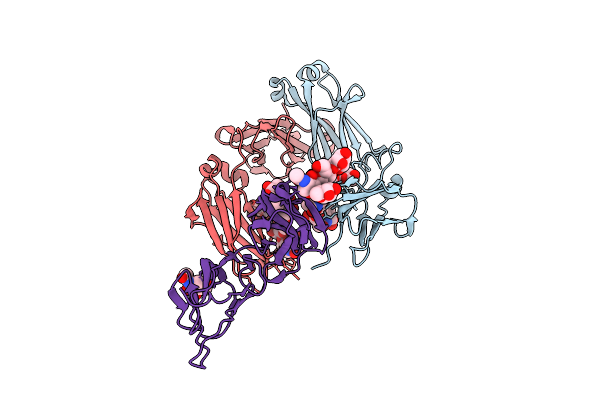 |
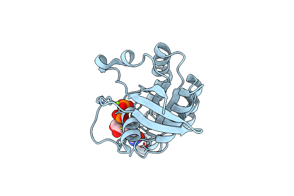
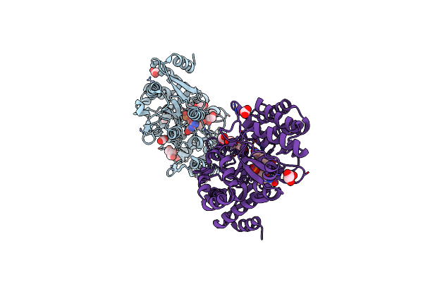
Crystal Structure Of Human Igg1 Fc Fragment-Fc-Gamma Receptor Iia Complex H131 Variant
Organism: Homo sapiens
Method: X-RAY DIFFRACTION Release Date: 2025-12-03 Classification: IMMUNE SYSTEM Ligands: NAG |
 |
Crystal Structure Of Human Igg1 Fc Fragment-Fc-Gamma Receptor Iia Complex R131 Variant
Organism: Homo sapiens
Method: X-RAY DIFFRACTION Release Date: 2025-12-03 Classification: IMMUNE SYSTEM Ligands: NAG, MPD |
 |
Crystal Structure Of Human Igg2 Fc Fragment-Fc-Gamma Receptor Iia Complex H131 Variant
Organism: Homo sapiens
Method: X-RAY DIFFRACTION Release Date: 2025-12-03 Classification: IMMUNE SYSTEM Ligands: NAG |
 |
Organism: Homo sapiens
Method: X-RAY DIFFRACTION Release Date: 2025-12-03 Classification: IMMUNE SYSTEM Ligands: HOH |
 |
Organism: Severe acute respiratory syndrome coronavirus 2
Method: ELECTRON MICROSCOPY Release Date: 2025-10-01 Classification: VIRAL PROTEIN Ligands: NAG |
 |
Organism: Severe acute respiratory syndrome coronavirus 2
Method: ELECTRON MICROSCOPY Release Date: 2025-10-01 Classification: VIRAL PROTEIN Ligands: NAG |
 |
Organism: Severe acute respiratory syndrome coronavirus 2
Method: ELECTRON MICROSCOPY Release Date: 2025-10-01 Classification: VIRAL PROTEIN Ligands: NAG |
 |
Organism: Severe acute respiratory syndrome coronavirus 2
Method: ELECTRON MICROSCOPY Release Date: 2025-10-01 Classification: VIRAL PROTEIN Ligands: NAG |
 |
Organism: Homo sapiens
Method: X-RAY DIFFRACTION Release Date: 2025-07-09 Classification: PROTEIN BINDING Ligands: ACT, EDO |
 |
Organism: Homo sapiens
Method: X-RAY DIFFRACTION Resolution:2.10 Å Release Date: 2024-03-27 Classification: PROTEIN BINDING Ligands: ZTO |
 |
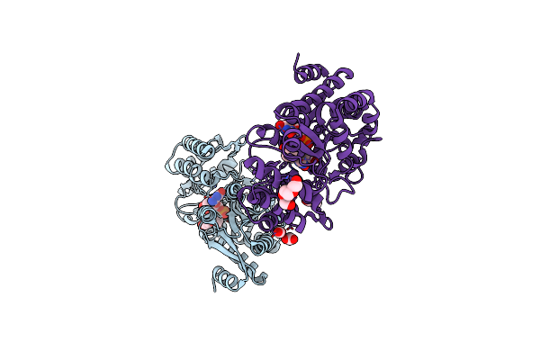
Organism: Homo sapiens
Method: X-RAY DIFFRACTION Resolution:1.40 Å Release Date: 2024-03-20 Classification: HYDROLASE Ligands: GDP, MG |
 |
Crystal Structure Of A Dinucleotide-Binding Protein Of Abc Transporter Endogenously Bound To Uridylyl-3'-5'-Phospho-Guanosine (Form I)
Organism: Thermus thermophilus hb8
Method: X-RAY DIFFRACTION Resolution:2.15 Å Release Date: 2021-03-10 Classification: TRANSPORT PROTEIN Ligands: CL, PO2, CO2, EDO, GOL, PEG, PDO, PG4, 1PE, FGO, PGO, PGR |
 |
Crystal Structure Of A Dinucleotide-Binding Protein Of Abc Transporter Endogenously Bound To Uridylyl-3'-5'-Phospho-Guanosine (Form Ii)
Organism: Thermus thermophilus hb8
Method: X-RAY DIFFRACTION Resolution:1.80 Å Release Date: 2021-03-10 Classification: TRANSPORT PROTEIN Ligands: CL, NA, CO2, PO2, PO4, EDO, PDO, GOL, BGQ, PG4, FGO, CO3, PO3 |
 |
Crystal Structure Of A Dinucleotide-Binding Protein Of Abc Transporter Endogenously Bound To Uridylyl-3'-5'-Phospho-Guanosine (Form Iii)
Organism: Thermus thermophilus hb8
Method: X-RAY DIFFRACTION Resolution:1.85 Å Release Date: 2021-03-10 Classification: TRANSPORT PROTEIN Ligands: CL, SO3, EDO, GOL, FGO, CO2, SO2 |
 |
Crystal Structure Of A Dinucleotide-Binding Protein (Y56F) Of Abc Transporter Endogenously Bound To Uridylyl-3'-5'-Phospho-Guanosine
Organism: Thermus thermophilus hb8
Method: X-RAY DIFFRACTION Resolution:2.00 Å Release Date: 2021-03-10 Classification: TRANSPORT PROTEIN Ligands: CO2, EDO, GOL, FGO, NA, CO3, PEG |
 |
Crystal Structure Of A Dinucleotide-Binding Protein (F79A) Of Abc Transporter Endogenously Bound To Uridylyl-3'-5'-Phospho-Guanosine (Form I)
Organism: Thermus thermophilus hb8
Method: X-RAY DIFFRACTION Resolution:1.77 Å Release Date: 2021-03-10 Classification: TRANSPORT PROTEIN Ligands: CL, CO2, EDO, GOL, FGO |
 |
Crystal Structure Of A Dinucleotide-Binding Protein (F79A) Of Abc Transporter Endogenously Bound To Uridylyl-3'-5'-Phospho-Guanosine (Form Ii)
Organism: Thermus thermophilus hb8
Method: X-RAY DIFFRACTION Resolution:1.85 Å Release Date: 2021-03-10 Classification: TRANSPORT PROTEIN Ligands: CL, EDO, FGO, CO2, CO3, PGR, PGE |