
Search Count: 191
 |
Organism: Homo sapiens
Method: ELECTRON MICROSCOPY Release Date: 2025-12-24 Classification: MEMBRANE PROTEIN Ligands: LMN, ZMA |
 |
Organism: Homo sapiens
Method: ELECTRON MICROSCOPY Release Date: 2025-12-24 Classification: MEMBRANE PROTEIN Ligands: LMN, ZMA |
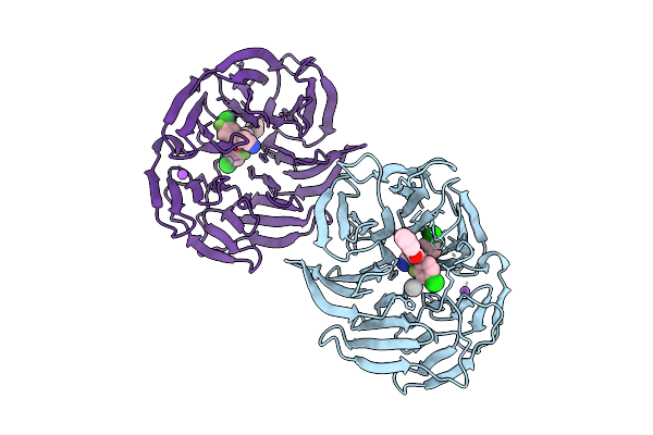 |
Organism: Homo sapiens
Method: X-RAY DIFFRACTION Release Date: 2025-11-26 Classification: TRANSPORT PROTEIN Ligands: A1CY9, NA, UNX |
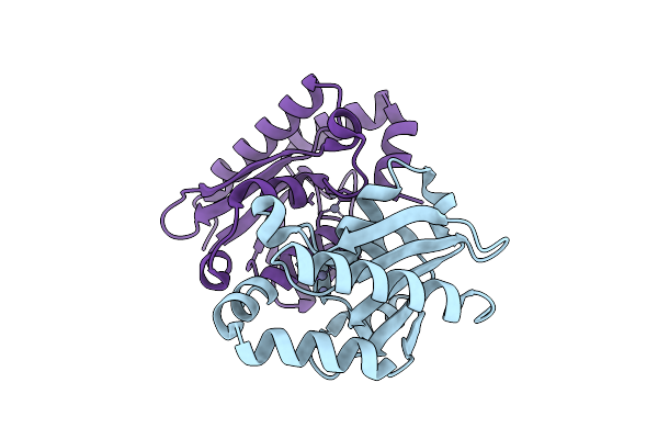 |
Organism: Saccharomyces cerevisiae (strain atcc 204508 / s288c)
Method: X-RAY DIFFRACTION Release Date: 2025-10-22 Classification: ANTITUMOR PROTEIN Ligands: ZN |
 |
Organism: Xenopus laevis, Homo sapiens
Method: ELECTRON MICROSCOPY Release Date: 2025-10-15 Classification: TRANSPORT PROTEIN |
 |
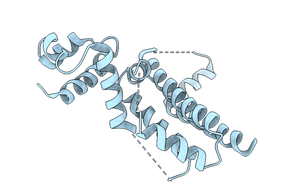
Structure Of Xenopus Kcnq1(E150R/R221E)-Cam With The Vsd In The Intermediate State
Organism: Xenopus laevis, Homo sapiens
Method: ELECTRON MICROSCOPY Release Date: 2025-10-15 Classification: TRANSPORT PROTEIN |
 |
Crystal Structure Of The Engineered Sulfonylurea Repressor Esr (L7-D1), Apo Form
Organism: Escherichia coli
Method: X-RAY DIFFRACTION Release Date: 2025-10-08 Classification: DNA BINDING PROTEIN |
 |
Crystal Structure Of The Engineered Sulfonylurea Repressor Esr (L11-C6), Bound To Ethametsulfuron-Methyl
Organism: Escherichia coli
Method: X-RAY DIFFRACTION Release Date: 2025-10-08 Classification: DNA BINDING PROTEIN Ligands: RXF |
 |
Crystal Structure Of The Engineered Sulfonylurea Repressor Csr (L4.2-20), Apo Form
Organism: Escherichia coli
Method: X-RAY DIFFRACTION Release Date: 2025-10-08 Classification: DNA BINDING PROTEIN |
 |
Crystal Structure Of The Engineered Sulfonylurea Repressor Csr (L4.2-20), Bound To Chlorsulfuron
Organism: Escherichia coli
Method: X-RAY DIFFRACTION Release Date: 2025-10-08 Classification: DNA BINDING PROTEIN Ligands: 1CS |
 |
Organism: Homo sapiens
Method: ELECTRON MICROSCOPY Release Date: 2025-09-24 Classification: CELL ADHESION Ligands: NAG, CA, MN |
 |
Organism: Homo sapiens
Method: ELECTRON MICROSCOPY Release Date: 2025-09-24 Classification: CELL ADHESION Ligands: CA |
 |
Organism: Homo sapiens, Mus musculus
Method: ELECTRON MICROSCOPY Release Date: 2025-09-24 Classification: CELL ADHESION Ligands: NAG, CA |
 |
Organism: Homo sapiens, Mus musculus
Method: ELECTRON MICROSCOPY Release Date: 2025-09-24 Classification: CELL ADHESION Ligands: NAG, CA |
 |
Organism: Homo sapiens
Method: ELECTRON MICROSCOPY Release Date: 2025-09-24 Classification: CELL ADHESION Ligands: NAG, CA, MN |
 |
Organism: Homo sapiens
Method: SOLUTION NMR Release Date: 2025-08-06 Classification: PROTEIN BINDING |
 |
Sars-Cov-2 Nsp1 Bound To The Rhinolophus Lepidus 40S Ribosomal Subunit (Local Refinement Of The 40S Body)
Organism: Severe acute respiratory syndrome coronavirus 2, Homo sapiens, Rhinolophus lepidus
Method: ELECTRON MICROSCOPY Release Date: 2025-06-11 Classification: RIBOSOME Ligands: MG, K, ZN |
 |
Sars-Cov-2 Nsp1 Bound To The Rhinolophus Lepidus 40S Ribosome (Local Refinement Of The 40S Head)
Organism: Rhinolophus lepidus
Method: ELECTRON MICROSCOPY Release Date: 2025-06-11 Classification: RIBOSOME Ligands: MG, ZN, K |
 |
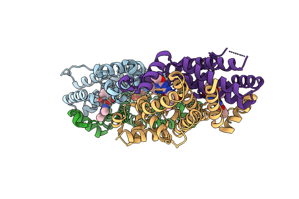
Organism: Bacillus thuringiensis
Method: X-RAY DIFFRACTION Resolution:3.11 Å Release Date: 2025-02-12 Classification: TOXIN |
 |
Organism: Homo sapiens, Rattus norvegicus
Method: ELECTRON MICROSCOPY Release Date: 2025-02-05 Classification: SIGNALING PROTEIN,CELL ADHESION Ligands: CA, NAG |