
Search Count: 109
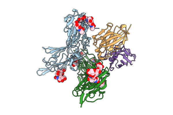 |
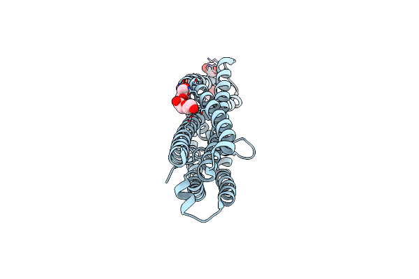
Organism: Plasmodium falciparum 3d7, Vicugna pacos
Method: X-RAY DIFFRACTION Release Date: 2025-07-30 Classification: PROTEIN BINDING Ligands: GOL, NAG, PEG |
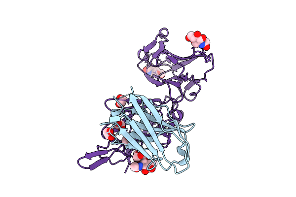 |
Organism: Vicugna pacos, Plasmodium falciparum 3d7
Method: X-RAY DIFFRACTION Release Date: 2025-07-30 Classification: PROTEIN BINDING Ligands: EDO, NAG |
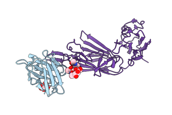 |
Organism: Vicugna pacos, Plasmodium falciparum 3d7
Method: X-RAY DIFFRACTION Release Date: 2025-07-30 Classification: PROTEIN BINDING Ligands: CIT |
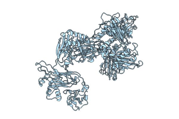 |
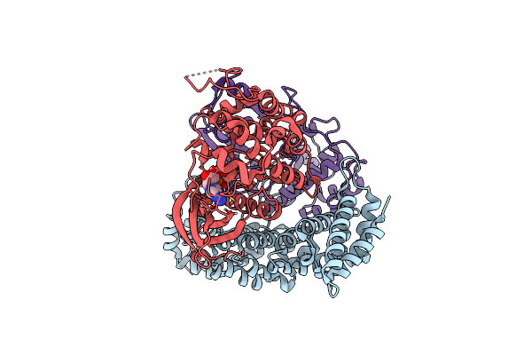
Organism: Plasmodium falciparum
Method: ELECTRON MICROSCOPY Release Date: 2025-07-30 Classification: PROTEIN BINDING |
 |
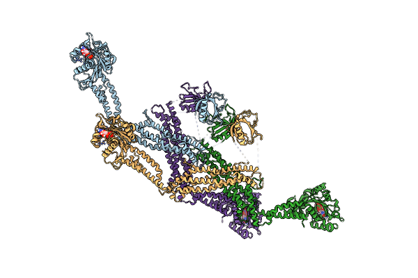
Organism: Plasmodium falciparum
Method: ELECTRON MICROSCOPY Release Date: 2025-07-30 Classification: PROTEIN BINDING |
 |
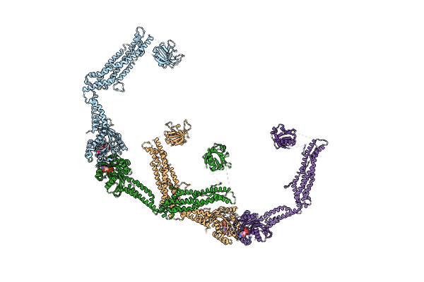
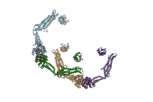
Structure Of The 50S Ribosomal Subunit From The Antibiotic-Producing Bacterium Streptomyces Fradiae
Organism: Streptomyces fradiae atcc 10745 = dsm 40063
Method: ELECTRON MICROSCOPY Release Date: 2025-05-14 Classification: RIBOSOME |
 |
Organism: Psychrobacter urativorans
Method: ELECTRON MICROSCOPY Release Date: 2025-03-26 Classification: RIBOSOME |
 |
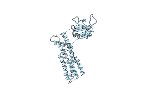
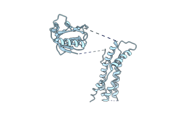
Organism: Plasmodium vivax
Method: X-RAY DIFFRACTION Resolution:2.40 Å Release Date: 2025-02-26 Classification: CELL ADHESION Ligands: MPD, GOL, EPE, PEG |
 |
Organism: Plasmodium vivax
Method: X-RAY DIFFRACTION Resolution:1.85 Å Release Date: 2025-02-26 Classification: CELL ADHESION Ligands: GOL, PEG |
 |
Structure Of The P1B7 Antibody Bound To The Sotorasib-Modified Kras G12C Peptide Presented By The A*03:01 Mhc I Complex
Organism: Homo sapiens
Method: ELECTRON MICROSCOPY Release Date: 2024-10-16 Classification: IMMUNE SYSTEM Ligands: MOV |
 |
Organism: Homo sapiens
Method: ELECTRON MICROSCOPY Release Date: 2024-07-10 Classification: TRANSFERASE Ligands: ADP |
 |
Cryo-Em Of The Gdp-Bound Human Dynamin (Full-Length) Polymer Assembled On The Membrane In The Super Constricted State
Organism: Homo sapiens
Method: ELECTRON MICROSCOPY Release Date: 2024-06-12 Classification: HYDROLASE Ligands: GDP, MG |
 |
Cryo-Em Of The Gdp-Bound Human Dynamin Polymer Assembled On The Membrane In The Super Constricted State (Tetramer Model)
Organism: Homo sapiens
Method: ELECTRON MICROSCOPY Release Date: 2024-06-12 Classification: HYDROLASE Ligands: GDP, MG |
 |
Cryo-Em Of The Gdp-Bound Human Dynamin Polymer Assembled On The Membrane In The Super Constricted State
Organism: Homo sapiens
Method: ELECTRON MICROSCOPY Release Date: 2024-05-01 Classification: HYDROLASE Ligands: GDP, MG |
 |
Cryo-Em Of The Gdp-Bound Human Dynamin Polymer Assembled On The Membrane In The Super Constricted State Showing The Ph Domain
Organism: Homo sapiens
Method: ELECTRON MICROSCOPY Release Date: 2024-05-01 Classification: HYDROLASE |
 |
Cryo-Em Of The Gdp-Bound Human Dynamin Polymer Assembled On The Membrane In The Super Constricted State Showing The Second Ph Domain
Organism: Homo sapiens
Method: ELECTRON MICROSCOPY Release Date: 2024-05-01 Classification: HYDROLASE |
 |
Cryo-Em Of The Gdp-Bound Human Dynamin Polymer Assembled On The Membrane In The Super Constricted State (Full Helix)
Organism: Homo sapiens
Method: ELECTRON MICROSCOPY Release Date: 2024-05-01 Classification: HYDROLASE Ligands: GDP, MG |
 |
Cryo-Em Of The Gdp-Bound Human Dynamin (Full-Length) Polymer Assembled On The Membrane In The Super Constricted State
Organism: Homo sapiens
Method: ELECTRON MICROSCOPY Release Date: 2024-05-01 Classification: HYDROLASE Ligands: GDP, MG |
 |
Cryo-Em Of The Gdp-Bound Human Dynamin (Full-Length) Polymer Assembled On The Membrane In The Super Constricted State (Full Helix)
Organism: Homo sapiens
Method: ELECTRON MICROSCOPY Release Date: 2024-05-01 Classification: HYDROLASE Ligands: GDP, MG |
 |
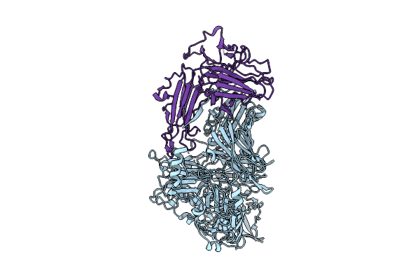
Organism: Human coronavirus oc43, Vicugna pacos
Method: X-RAY DIFFRACTION Resolution:2.90 Å Release Date: 2024-05-01 Classification: VIRAL PROTEIN Ligands: GOL |