
Search Count: 25
 |
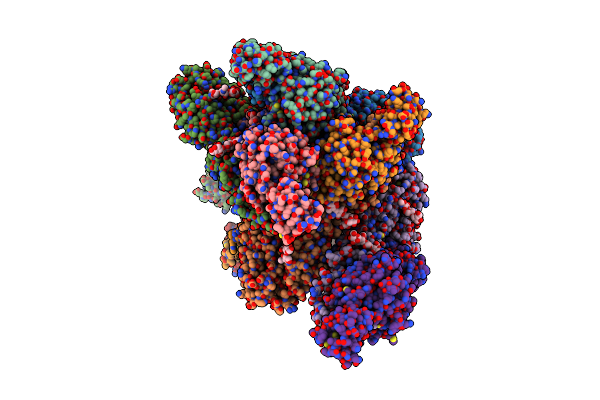
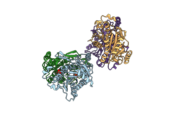
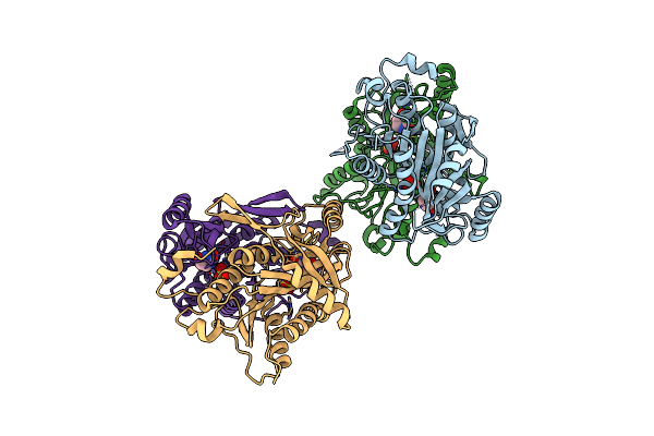
Organism: Mus musculus
Method: ELECTRON MICROSCOPY Release Date: 2023-09-20 Classification: HYDROLASE Ligands: PO4 |
 |
 |
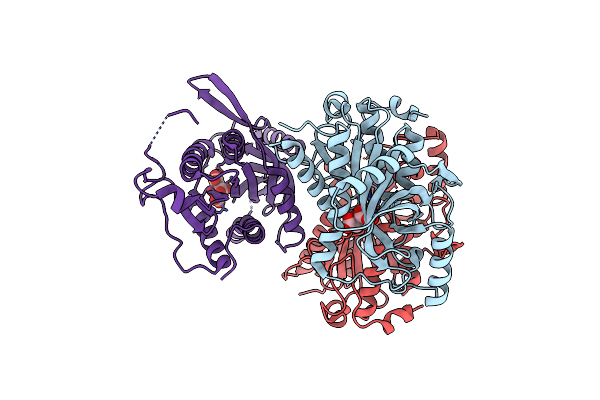
Em Structure Of The Dna Wrapping In Bacterial Open Transcription Initiation Complex
Organism: Enterobacteria phage lambda, Escherichia coli k-12
Method: ELECTRON MICROSCOPY Release Date: 2020-05-27 Classification: transcription/dna |
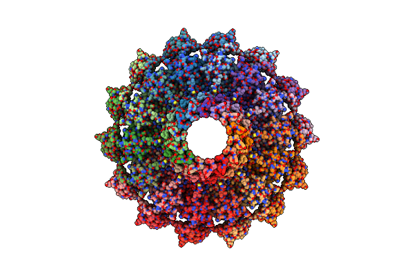 |
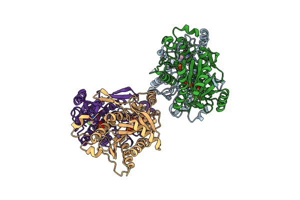
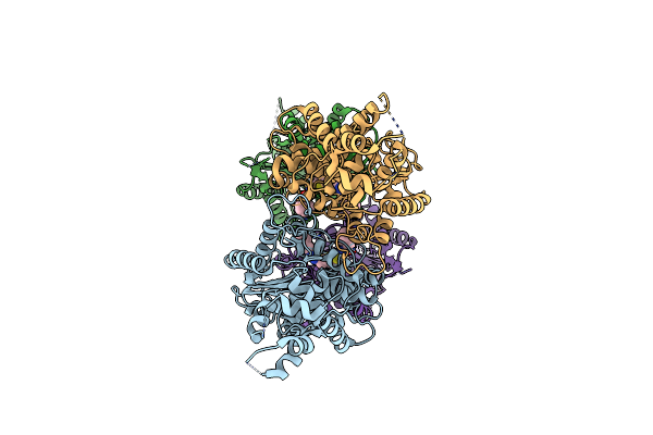
Cryo-Em Structure Of The Bacteria-Killing Type Iv Secretion System Core Complex From Xanthomonas Citri
Organism: Xanthomonas axonopodis pv. citri (strain 306)
Method: ELECTRON MICROSCOPY Release Date: 2018-10-24 Classification: MEMBRANE PROTEIN |
 |
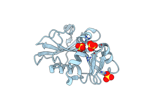
Crystal Structure Of Isoform 2 Of Uridine Phosphorylase From Schistosoma Mansoni In Complex With Citrate
Organism: Schistosoma mansoni
Method: X-RAY DIFFRACTION Resolution:1.98 Å Release Date: 2016-03-16 Classification: TRANSFERASE Ligands: FLC |
 |
Crystal Structure Of Isoform 2 Of Uridine Phosphorylase From Schistosoma Mansoni Apo Form
Organism: Schistosoma mansoni
Method: X-RAY DIFFRACTION Resolution:2.02 Å Release Date: 2016-03-16 Classification: TRANSFERASE |
 |
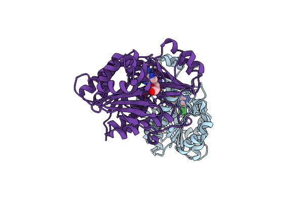
Crystal Structure Of Uridine Phosphorylase From Schistosoma Mansoni In Apo Form
Organism: Schistosoma mansoni
Method: X-RAY DIFFRACTION Resolution:1.89 Å Release Date: 2015-10-14 Classification: TRANSFERASE Ligands: SO4 |
 |
Crystal Structure Of Uridine Phosphorylase From Schistosoma Mansoni In Complex With Thymine
Organism: Schistosoma mansoni
Method: X-RAY DIFFRACTION Resolution:1.93 Å Release Date: 2015-10-14 Classification: TRANSFERASE Ligands: SO4, TDR |
 |
Crystal Structure Of Uridine Phosphorylase From Schistosoma Mansoni In Complex With 5-Fluorouracil
Organism: Schistosoma mansoni
Method: X-RAY DIFFRACTION Resolution:2.00 Å Release Date: 2015-10-14 Classification: TRANSFERASE Ligands: SO4, URF |
 |
Crystal Structure Of Uridine Phosphorylase From Schistosoma Mansoni In Complex With Thymidine
Organism: Schistosoma mansoni
Method: X-RAY DIFFRACTION Resolution:1.66 Å Release Date: 2015-07-15 Classification: TRANSFERASE Ligands: THM, SO4 |
 |
Crystal Structure Of Uridine Phosphorylase From Schistosoma Mansoni In Complex With Uracil
Organism: Schistosoma mansoni
Method: X-RAY DIFFRACTION Resolution:1.92 Å Release Date: 2015-07-15 Classification: TRANSFERASE Ligands: SO4, URA |
 |
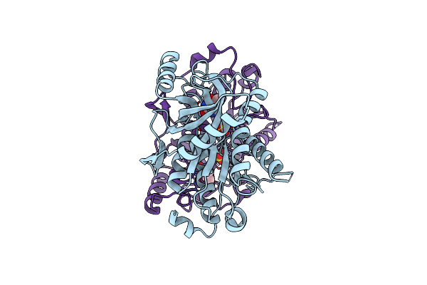
Organism: Mus musculus
Method: X-RAY DIFFRACTION Resolution:2.77 Å Release Date: 2013-08-14 Classification: HYDROLASE Ligands: 04A |
 |
Organism: Schistosoma mansoni
Method: X-RAY DIFFRACTION Resolution:1.95 Å Release Date: 2013-03-06 Classification: OXIDOREDUCTASE Ligands: SO4 |
 |
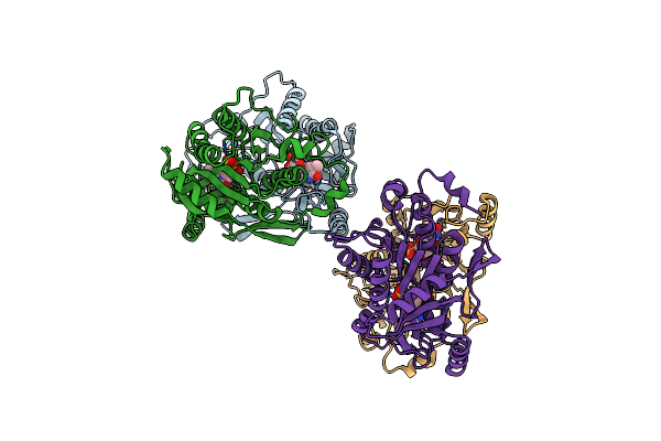
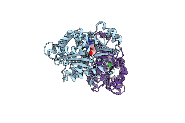
Adenosine Kinase From Schistosoma Mansoni In Complex With 2-Fluoroadenosine
Organism: Schistosoma mansoni
Method: X-RAY DIFFRACTION Resolution:2.40 Å Release Date: 2012-11-28 Classification: TRANSFERASE Ligands: ADN, CL, 2FA |
 |
Organism: Schistosoma mansoni
Method: X-RAY DIFFRACTION Resolution:2.44 Å Release Date: 2012-11-14 Classification: TRANSFERASE Ligands: ADN, CL |
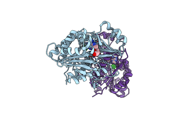 |
Adenosine Kinase From Schistosoma Mansoni In Complex With Adenosine In Occluded Loop Conformation
Organism: Schistosoma mansoni
Method: X-RAY DIFFRACTION Resolution:2.26 Å Release Date: 2012-11-14 Classification: TRANSFERASE Ligands: ADN, CL |
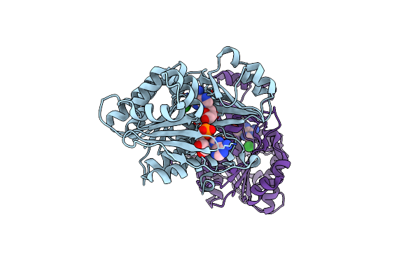 |
Adenosine Kinase From Schistosoma Mansoni In Complex With Adenosine And Amp
Organism: Schistosoma mansoni
Method: X-RAY DIFFRACTION Resolution:2.30 Å Release Date: 2012-10-31 Classification: TRANSFERASE Ligands: ADN, AMP, CL |
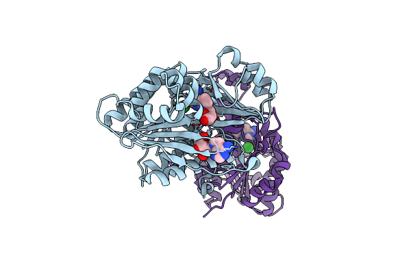 |
Organism: Schistosoma mansoni
Method: X-RAY DIFFRACTION Resolution:2.34 Å Release Date: 2012-10-31 Classification: TRANSFERASE Ligands: TBN, CL |
 |
Organism: Moniliophthora perniciosa
Method: X-RAY DIFFRACTION Resolution:1.34 Å Release Date: 2012-07-11 Classification: UNKNOWN FUNCTION Ligands: ACT, ZN, CL, NA |
 |
Organism: Moniliophthora perniciosa
Method: X-RAY DIFFRACTION Resolution:1.34 Å Release Date: 2012-07-11 Classification: UNKNOWN FUNCTION |