
Search Count: 28
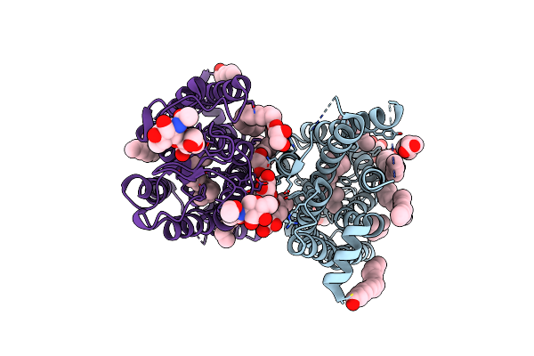 |
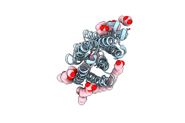
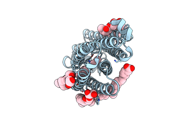
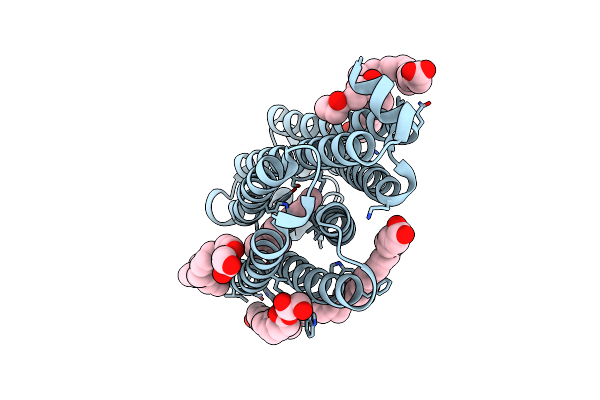
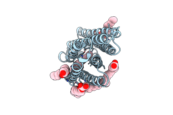
Dark State Crystal Structure Of Bovine Rhodopsin In Lipidic Cubic Phase (Sacla)
Organism: Bos taurus
Method: X-RAY DIFFRACTION Resolution:1.80 Å Release Date: 2023-03-29 Classification: MEMBRANE PROTEIN Ligands: ACE, RET, NAG, DAO, OLC, PLM |
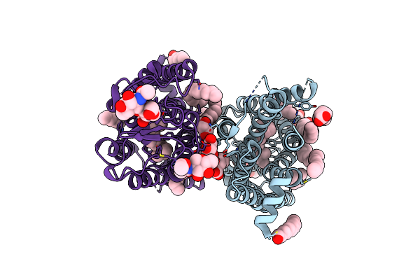 |
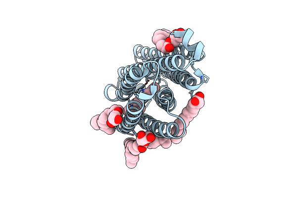
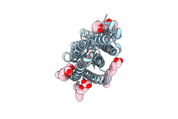
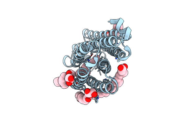
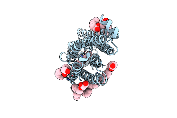
Dark State Crystal Structure Of Bovine Rhodopsin In Lipidic Cubic Phase (Swissfel)
Organism: Bos taurus
Method: X-RAY DIFFRACTION Resolution:1.80 Å Release Date: 2023-03-29 Classification: MEMBRANE PROTEIN Ligands: ACE, RET, NAG, DAO, OLC, PLM |
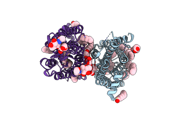 |
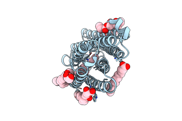
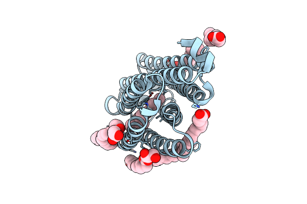
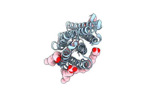
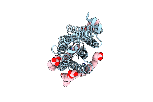
1 Picosecond Light Activated Crystal Structure Of Bovine Rhodopsin In Lipidic Cubic Phase
Organism: Bos taurus
Method: X-RAY DIFFRACTION Resolution:1.80 Å Release Date: 2023-03-29 Classification: MEMBRANE PROTEIN Ligands: ACE, RET, NAG, DAO, OLC, PLM |
 |
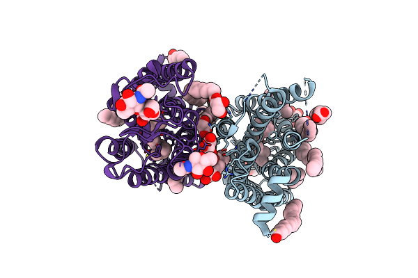
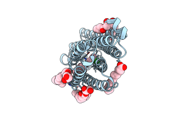
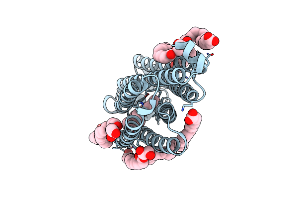
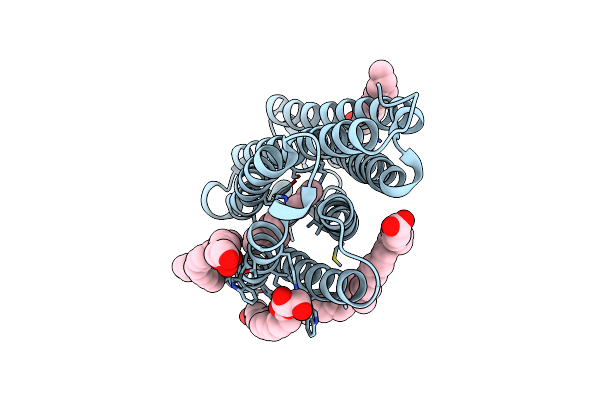
10 Picosecond Light Activated Crystal Structure Of Bovine Rhodopsin In Lipidic Cubic Phase
Organism: Bos taurus
Method: X-RAY DIFFRACTION Resolution:1.80 Å Release Date: 2023-03-29 Classification: MEMBRANE PROTEIN Ligands: ACE, RET, NAG, DAO, OLC, PLM |
 |
100 Picosecond Light Activated Crystal Structure Of Bovine Rhodopsin In Lipidic Cubic Phase (Sacla)
Organism: Bos taurus
Method: X-RAY DIFFRACTION Resolution:1.80 Å Release Date: 2023-03-29 Classification: MEMBRANE PROTEIN Ligands: RET, NAG, DAO, OLC, PLM |
 |
Organism: Nonlabens marinus s1-08
Method: X-RAY DIFFRACTION Resolution:1.45 Å Release Date: 2022-02-09 Classification: MEMBRANE PROTEIN Ligands: RET, CL, OLC, OLA |
 |
Nmhr Light State Structure At 10 Ps After Photoexcitation Determined By Serial Femtosecond Crystallography (With Extrapolated, Dark And Light Dataset)
Organism: Nonlabens marinus s1-08
Method: X-RAY DIFFRACTION Resolution:1.90 Å Release Date: 2022-02-09 Classification: MEMBRANE PROTEIN Ligands: RET, CL, OLC, OLA |
 |
Nmhr Light State Structure At 10 Ns After Photoexcitation Determined By Serial Femtosecond Crystallography (With Extrapolated, Dark And Light Dataset)
Organism: Nonlabens marinus s1-08
Method: X-RAY DIFFRACTION Resolution:1.80 Å Release Date: 2022-02-09 Classification: MEMBRANE PROTEIN Ligands: RET, CL, OLC, OLA |
 |
Nmhr Light State Structure At 1 Us After Photoexcitation Determined By Serial Femtosecond Crystallography (With Extrapolated, Dark And Light Dataset)
Organism: Nonlabens marinus s1-08
Method: X-RAY DIFFRACTION Resolution:1.80 Å Release Date: 2022-02-09 Classification: MEMBRANE PROTEIN Ligands: RET, CL, OLC, OLA |
 |
Nmhr Light State Structure At 20 Us After Photoexcitation Determined By Serial Femtosecond Crystallography (With Extrapolated, Dark And Light Dataset)
Organism: Nonlabens marinus s1-08
Method: X-RAY DIFFRACTION Resolution:1.80 Å Release Date: 2022-02-09 Classification: MEMBRANE PROTEIN Ligands: RET, CL, OLC, OLA |
 |
Nmhr Light State Structure At 300 Us After Photoexcitation Determined By Serial Femtosecond Crystallography (With Extrapolated, Dark And Light Dataset)
Organism: Nonlabens marinus s1-08
Method: X-RAY DIFFRACTION Resolution:1.90 Å Release Date: 2022-02-09 Classification: MEMBRANE PROTEIN Ligands: RET, OLC, OLA |
 |
Organism: Nonlabens marinus s1-08
Method: X-RAY DIFFRACTION Resolution:1.80 Å Release Date: 2022-02-09 Classification: MEMBRANE PROTEIN Ligands: RET, CL, OLC, OLA |
 |
Nmhr Light State Structure At 2.5 Ms (0 - 5 Ms) After Photoexcitation Determined By Serial Millisecond Crystallography
Organism: Nonlabens marinus s1-08
Method: X-RAY DIFFRACTION Resolution:2.20 Å Release Date: 2022-02-09 Classification: MEMBRANE PROTEIN Ligands: RET, OLC, OLA |
 |
Nmhr Light State Structure At 7.5 Ms (5 - 10 Ms) After Photoexcitation Determined By Serial Millisecond Crystallography
Organism: Nonlabens marinus s1-08
Method: X-RAY DIFFRACTION Resolution:2.10 Å Release Date: 2022-02-09 Classification: MEMBRANE PROTEIN Ligands: RET, OLC, OLA |
 |
Nmhr Light State Structure At 12.5 Ms (10 - 15 Ms) After Photoexcitation Determined By Serial Millisecond Crystallography
Organism: Nonlabens marinus s1-08
Method: X-RAY DIFFRACTION Resolution:2.20 Å Release Date: 2022-02-09 Classification: MEMBRANE PROTEIN Ligands: RET, OLC, OLA |
 |
Nmhr Light State Structure At 17.5 Ms (15 - 20 Ms) After Photoexcitation Determined By Serial Millisecond Crystallography
Organism: Nonlabens marinus s1-08
Method: X-RAY DIFFRACTION Resolution:2.40 Å Release Date: 2022-02-09 Classification: MEMBRANE PROTEIN Ligands: RET, OLC, OLA |
 |
Nmhr Light State Structure At 22.5 Ms (20 - 25 Ms) After Photoexcitation Determined By Serial Millisecond Crystallography
Organism: Nonlabens marinus s1-08
Method: X-RAY DIFFRACTION Resolution:2.60 Å Release Date: 2022-02-09 Classification: MEMBRANE PROTEIN Ligands: RET, CL, OLC, OLA |
 |
Nmhr Light State Structure At 27.5 Ms (25 - 30 Ms) After Photoexcitation Determined By Serial Millisecond Crystallography
Organism: Nonlabens marinus s1-08
Method: X-RAY DIFFRACTION Resolution:2.70 Å Release Date: 2022-02-09 Classification: MEMBRANE PROTEIN Ligands: RET, CL, OLC, OLA |
 |
Nmhr Light State Structure At 32.5 Ms (30 - 35 Ms) After Photoexcitation Determined By Serial Millisecond Crystallography
Organism: Nonlabens marinus s1-08
Method: X-RAY DIFFRACTION Resolution:2.50 Å Release Date: 2022-02-09 Classification: MEMBRANE PROTEIN Ligands: RET, CL, OLC, OLA |
 |
Nmhr Light State Structure At 37.5 Ms (35 - 40 Ms) After Photoexcitation Determined By Serial Millisecond Crystallography
Organism: Nonlabens marinus s1-08
Method: X-RAY DIFFRACTION Resolution:2.50 Å Release Date: 2022-02-09 Classification: MEMBRANE PROTEIN Ligands: RET, CL, OLC, OLA |