
Search Count: 54
 |
Organism: Arundo donax
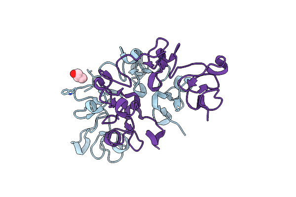
Method: X-RAY DIFFRACTION Resolution:1.70 Å Release Date: 2021-07-14 Classification: SUGAR BINDING PROTEIN Ligands: GOL |
 |
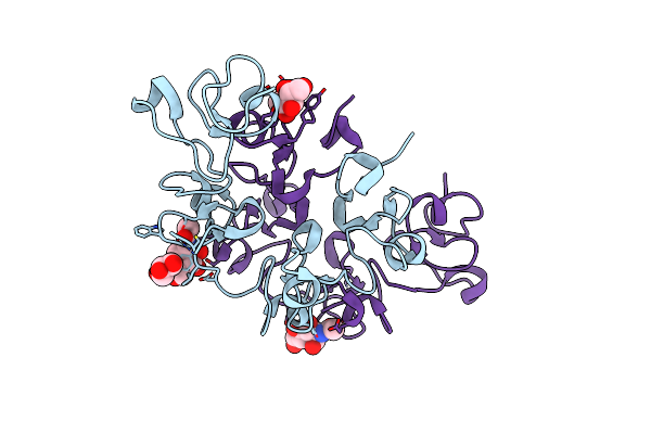
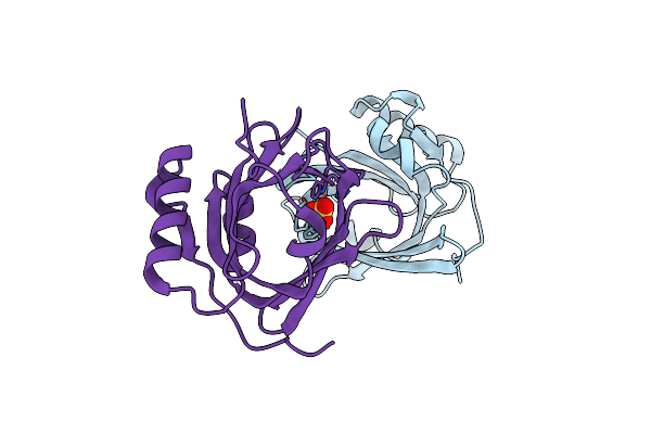
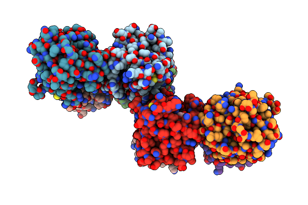
Three Dimensional Structure Of The Giant Reed (Arundodonax) Lectin (Adl) Complex With N-Acetyl Glucosamine
Organism: Arundo donax
Method: X-RAY DIFFRACTION Resolution:1.75 Å Release Date: 2021-07-14 Classification: SUGAR BINDING PROTEIN Ligands: NAG, GOL |
 |
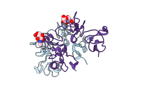
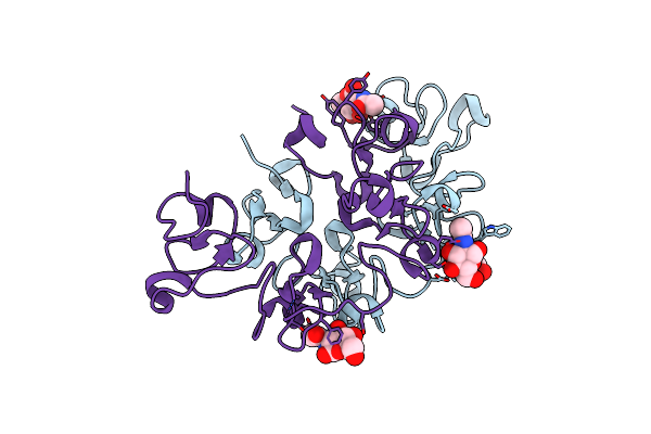
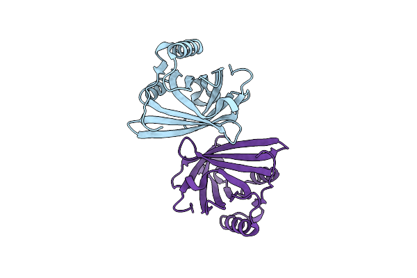
Three Dimensional Structure Of The Giant Reed (Arundodonax) Lectin (Adl) Complex With N-Acetyl Lactosamine
Organism: Arundo donax
Method: X-RAY DIFFRACTION Resolution:1.50 Å Release Date: 2021-07-14 Classification: SUGAR BINDING PROTEIN Ligands: NAG, GOL |
 |
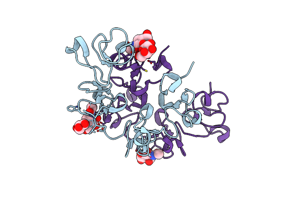
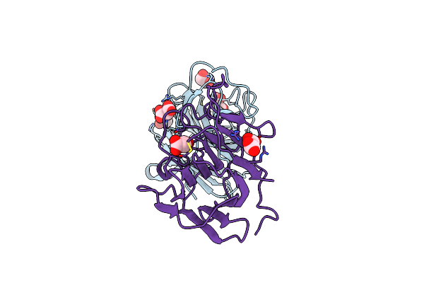
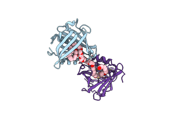
Three Dimensional Structure Of The Giant Reed (Arundodonax) Lectin (Adl) Complex With Sialic Acid
Organism: Arundo donax
Method: X-RAY DIFFRACTION Resolution:1.60 Å Release Date: 2021-07-14 Classification: SUGAR BINDING PROTEIN Ligands: SIA, GOL |
 |
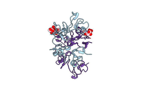
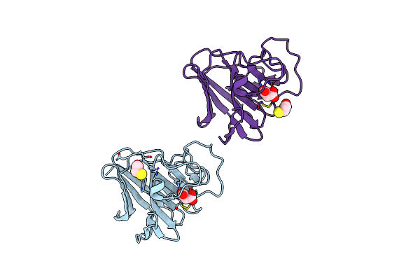
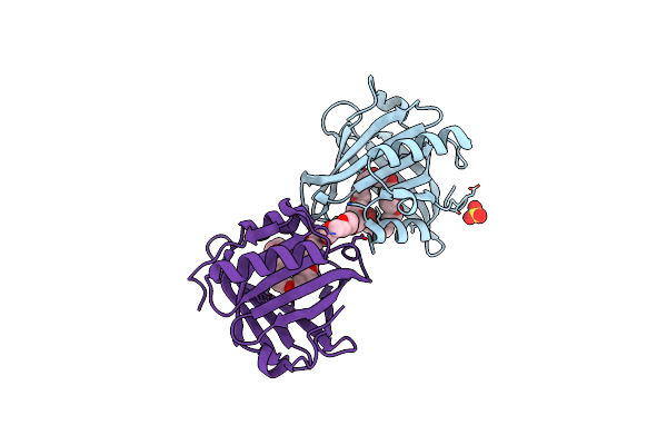
Three Dimensional Structure Of The Giant Reed (Arundodonax) Lectin (Adl) Complex With N,N'-Diacetylchitobiose; 30 Seconds Soaking
Organism: Arundo donax
Method: X-RAY DIFFRACTION Resolution:1.50 Å Release Date: 2021-07-14 Classification: SUGAR BINDING PROTEIN Ligands: NAG, GOL |
 |
Three Dimensional Structure Of The Giant Reed (Arundodonax) Lectin (Adl) Complex With N,N'-Diacetylchitobiose; 60 Seconds Soaking
Organism: Arundo donax
Method: X-RAY DIFFRACTION Resolution:1.80 Å Release Date: 2021-07-14 Classification: SUGAR BINDING PROTEIN Ligands: NAG, GOL |
 |
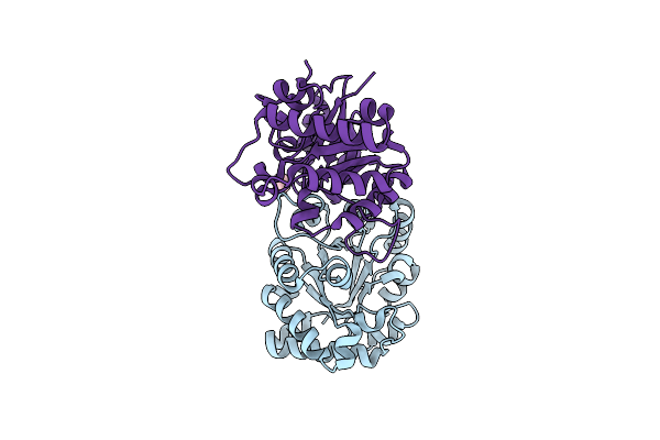
Organism: Homo sapiens
Method: X-RAY DIFFRACTION Resolution:1.60 Å Release Date: 2021-04-14 Classification: LIGASE Ligands: MLI, GOL |
 |
Organism: Homo sapiens
Method: X-RAY DIFFRACTION Resolution:1.80 Å Release Date: 2021-04-14 Classification: LIGASE Ligands: MLI, BME |
 |
Organism: Homo sapiens
Method: X-RAY DIFFRACTION Resolution:2.04 Å Release Date: 2018-05-16 Classification: ISOMERASE Ligands: IPA |
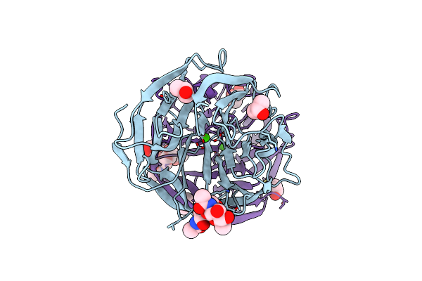 |
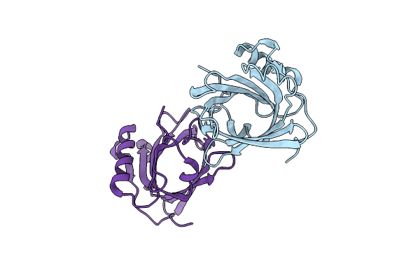
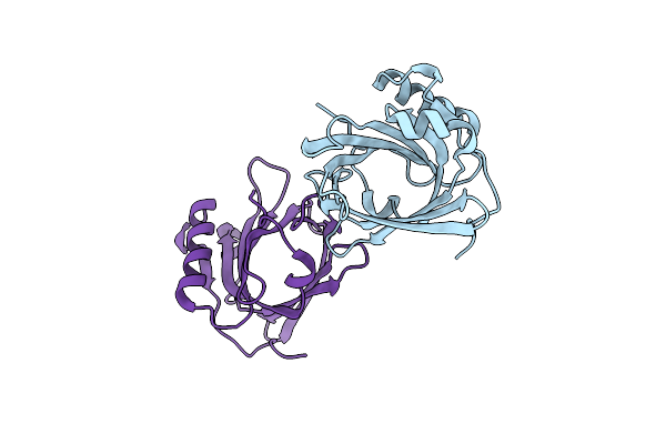
Organism: Cyprinus carpio
Method: X-RAY DIFFRACTION Resolution:1.35 Å Release Date: 2015-04-08 Classification: SUGAR BINDING PROTEIN Ligands: CA, IPA, GOL |
 |
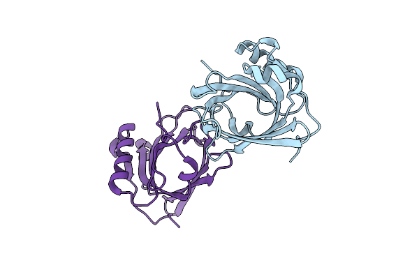
Organism: Cyprinus carpio
Method: X-RAY DIFFRACTION Resolution:1.70 Å Release Date: 2015-04-08 Classification: SUGAR BINDING PROTEIN Ligands: CA, NDG, 1PE, MG, NAG |
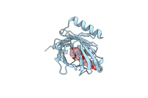 |
Threedimensional Structure Of The C65A Mutant Of Human Lipocalin-Type Prostaglandin D Synthase Olo-Form
Organism: Homo sapiens
Method: X-RAY DIFFRACTION Resolution:1.40 Å Release Date: 2014-08-06 Classification: ISOMERASE Ligands: PE3, SO4 |
 |
Three-Dimensional Structure Of The C65A Mutant Of Human Lipocalin-Type Prostaglandin D Synthase Apo-Form
Organism: Homo sapiens
Method: X-RAY DIFFRACTION Resolution:1.40 Å Release Date: 2014-08-06 Classification: ISOMERASE Ligands: SO4 |
 |
Three-Dimensional Structure Of The C65A Mutant Of Human Lipocalin-Type Prostaglandin D Synthase Holo-Form Second Space Group
Organism: Homo sapiens
Method: X-RAY DIFFRACTION Resolution:1.55 Å Release Date: 2014-08-06 Classification: ISOMERASE Ligands: PEU |
 |
Three-Dimensional Structure Of The C65A-K59A Double Mutant Of Human Lipocalin-Type Prostaglandin D Synthase Apo-Form
Organism: Homo sapiens
Method: X-RAY DIFFRACTION Resolution:1.66 Å Release Date: 2014-08-06 Classification: ISOMERASE |
 |
Three-Dimensional Structure Of The C65A-K59A Double Mutant Of Human Lipocalin-Type Prostaglandin D Synthase Holo-Form
Organism: Homo sapiens
Method: X-RAY DIFFRACTION Resolution:1.60 Å Release Date: 2014-08-06 Classification: ISOMERASE Ligands: SO4, PEU |
 |
Three-Dimensional Structure Of The C65A-K59A Double Mutant Of Human Lipocalin-Type Prostaglandin D Synthase Holo, Second Crystal Form
Organism: Homo sapiens
Method: X-RAY DIFFRACTION Resolution:1.80 Å Release Date: 2014-08-06 Classification: ISOMERASE Ligands: PEU |
 |
Three-Dimensional Structure Of The C65A-W54F Double Mutant Of Human Lipocalin-Type Prostaglandin D Synthase Apo-Form
Organism: Homo sapiens
Method: X-RAY DIFFRACTION Resolution:1.75 Å Release Date: 2014-08-06 Classification: ISOMERASE |
 |
Three-Dimensional Structure Of The C65A-W112F Double Mutant Of Human Lipocalin-Type Prostaglandin D Synthase Apo-Form
Organism: Homo sapiens
Method: X-RAY DIFFRACTION Resolution:1.40 Å Release Date: 2014-08-06 Classification: ISOMERASE |
 |
Three-Dimensional Structure Of The C65A-W54F-W112F Triple Mutant Of Human Lipocalin-Type Prostaglandin D Synthase Apo-Form
Organism: Homo sapiens
Method: X-RAY DIFFRACTION Resolution:1.69 Å Release Date: 2014-08-06 Classification: ISOMERASE |