
Search Count: 6
 |
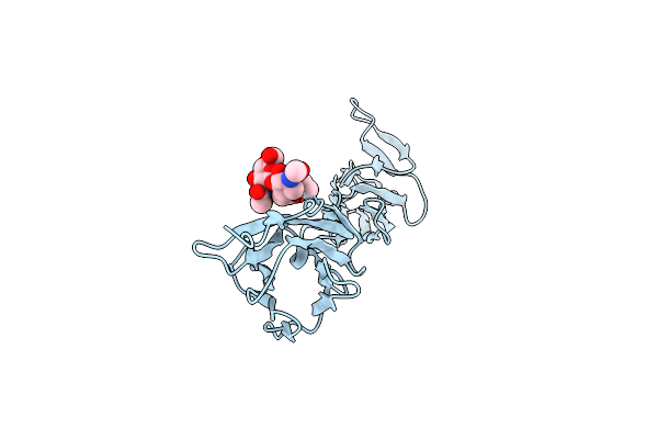
Organism: Drosophila melanogaster
Method: X-RAY DIFFRACTION Resolution:1.80 Å Release Date: 2019-02-06 Classification: CELL ADHESION Ligands: GOL, NAG |
 |
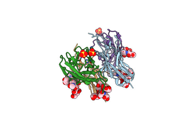
Organism: Drosophila melanogaster
Method: X-RAY DIFFRACTION Resolution:2.50 Å Release Date: 2019-02-06 Classification: CELL ADHESION Ligands: SO4, GOL |
 |
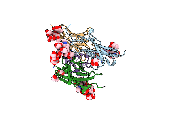
Organism: Drosophila melanogaster
Method: X-RAY DIFFRACTION Resolution:2.40 Å Release Date: 2019-02-06 Classification: CELL ADHESION Ligands: SO4, NAG |
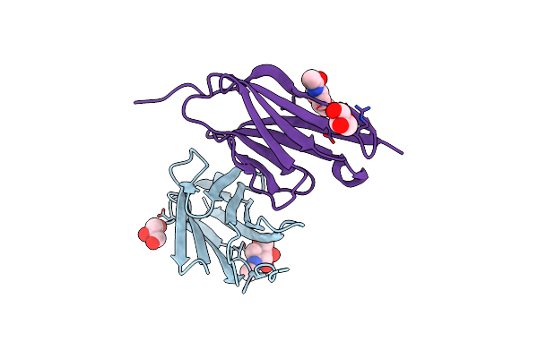 |
Organism: Drosophila melanogaster
Method: X-RAY DIFFRACTION Resolution:1.90 Å Release Date: 2019-02-06 Classification: CELL ADHESION Ligands: NAG, GOL |
 |
Organism: Drosophila melanogaster
Method: X-RAY DIFFRACTION Resolution:1.85 Å Release Date: 2019-02-06 Classification: CELL ADHESION |
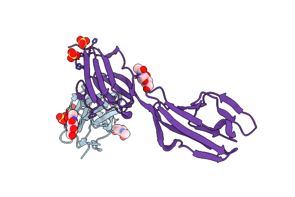 |
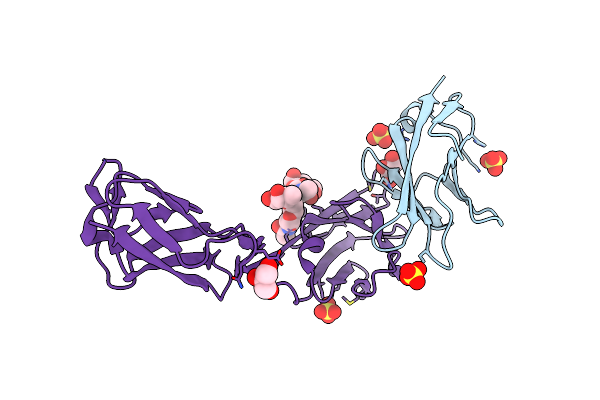
Crystal Structure Of The Complex Of Dpr6 Domain 1 Bound To Dip-Alpha Domain 1+2
Organism: Drosophila melanogaster
Method: X-RAY DIFFRACTION Resolution:2.30 Å Release Date: 2016-01-06 Classification: CELL ADHESION Ligands: NAG, SO4 |