
Search Count: 244
 |
Organism: Escherichia coli
Method: X-RAY DIFFRACTION Release Date: 2025-10-29 Classification: METAL BINDING PROTEIN Ligands: ZN |
 |
Organism: Escherichia coli
Method: X-RAY DIFFRACTION Release Date: 2025-10-29 Classification: METAL BINDING PROTEIN Ligands: ZN |
 |
Organism: Escherichia coli
Method: X-RAY DIFFRACTION Release Date: 2025-10-29 Classification: METAL BINDING PROTEIN Ligands: NI, NO3 |
 |
Organism: Escherichia coli
Method: X-RAY DIFFRACTION Release Date: 2025-10-29 Classification: METAL BINDING PROTEIN Ligands: CA, TRS |
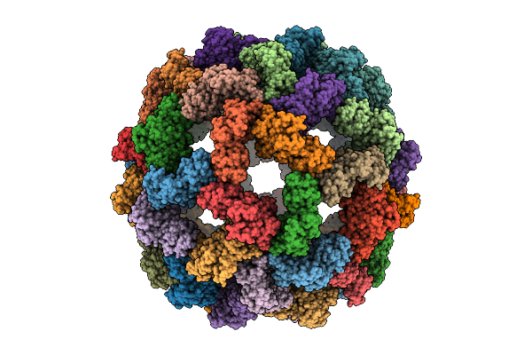 |
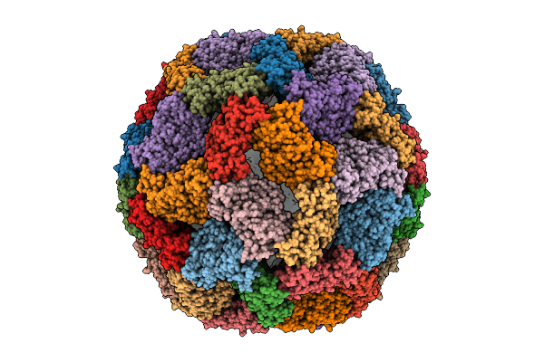
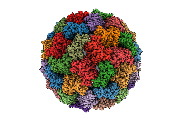
Cryo-Em Structure Of Designed Zinc-Induced Icosahedron Cage-I53-Zn1-Hehe-14
Organism: Escherichia coli
Method: ELECTRON MICROSCOPY Release Date: 2025-10-29 Classification: METAL BINDING PROTEIN Ligands: ZN |
 |
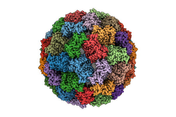
Cryo-Em Structure Of Designed Zinc-Induced Icosahedron Cage-I53-Zn1-Hehe-16
Organism: Escherichia coli
Method: ELECTRON MICROSCOPY Release Date: 2025-10-29 Classification: METAL BINDING PROTEIN Ligands: ZN |
 |
Organism: Escherichia coli
Method: ELECTRON MICROSCOPY Release Date: 2025-10-29 Classification: METAL BINDING PROTEIN Ligands: ZN |
 |
Organism: Escherichia coli
Method: ELECTRON MICROSCOPY Release Date: 2025-10-29 Classification: METAL BINDING PROTEIN Ligands: ZN |
 |
Organism: Escherichia coli
Method: ELECTRON MICROSCOPY Release Date: 2025-10-29 Classification: METAL BINDING PROTEIN Ligands: ZN |
 |
Organism: Escherichia coli
Method: ELECTRON MICROSCOPY Release Date: 2025-10-29 Classification: METAL BINDING PROTEIN Ligands: ZN |
 |
Organism: Escherichia coli
Method: ELECTRON MICROSCOPY Release Date: 2025-10-29 Classification: METAL BINDING PROTEIN Ligands: ZN |
 |
Organism: Escherichia coli
Method: ELECTRON MICROSCOPY Release Date: 2025-10-29 Classification: METAL BINDING PROTEIN Ligands: ZN |
 |
Organism: Escherichia coli
Method: ELECTRON MICROSCOPY Release Date: 2025-10-29 Classification: METAL BINDING PROTEIN Ligands: ZN |
 |
Organism: Escherichia coli
Method: ELECTRON MICROSCOPY Release Date: 2025-10-29 Classification: METAL BINDING PROTEIN Ligands: ZN |
 |
Organism: Escherichia coli
Method: ELECTRON MICROSCOPY Release Date: 2025-10-29 Classification: METAL BINDING PROTEIN Ligands: ZN |
 |
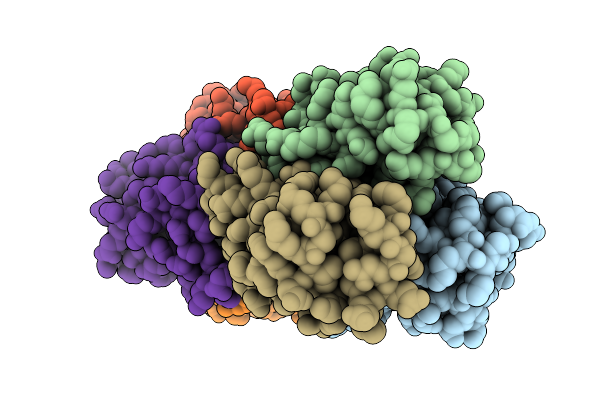
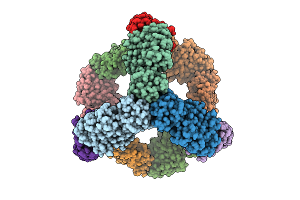
Cryo-Em Structure Of Designed Zinc-Induced Tetrahedron Cage-T32-Zn1-Hehe-34
Organism: Escherichia coli
Method: ELECTRON MICROSCOPY Release Date: 2025-10-29 Classification: METAL BINDING PROTEIN Ligands: ZN |
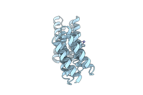 |
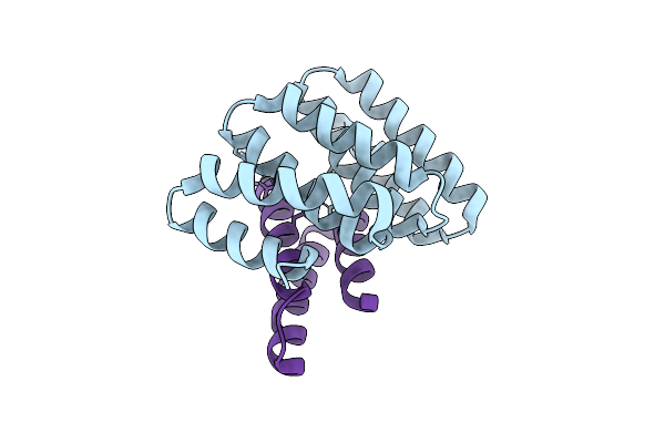
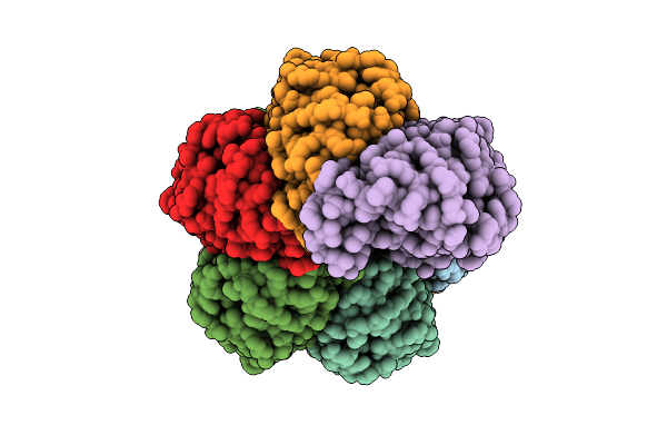
Cryo-Em Structure Of Monomer Of Designed Zinc-Induced Tetrahedron Cage-T32-Zn1-Hehe-34
Organism: Escherichia coli
Method: ELECTRON MICROSCOPY Release Date: 2025-10-29 Classification: METAL BINDING PROTEIN Ligands: ZN |
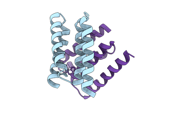 |
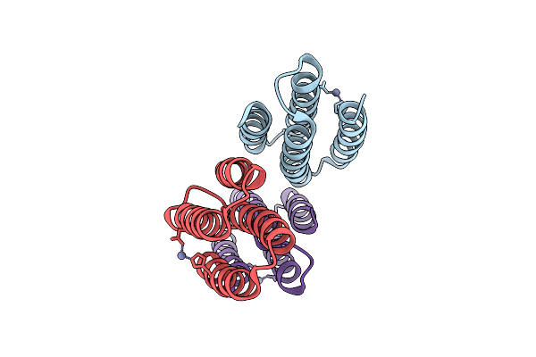
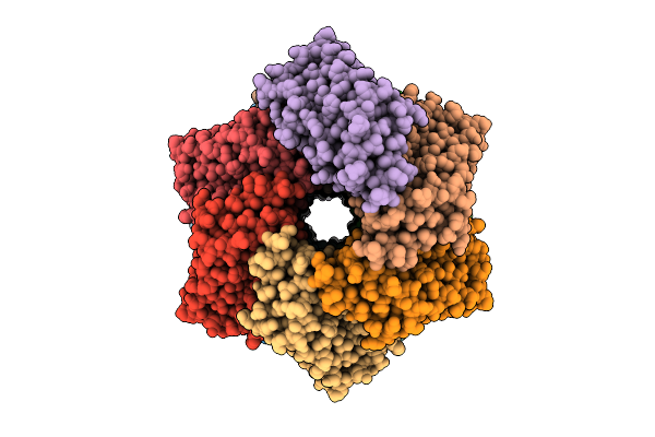
Cryo-Em Structure Of C2 Symmetric Interface Of Designed Zinc-Induced Tetrahedron Cage-T32-Zn1-Hehe-34
Organism: Escherichia coli
Method: ELECTRON MICROSCOPY Release Date: 2025-10-29 Classification: METAL BINDING PROTEIN Ligands: ZN |
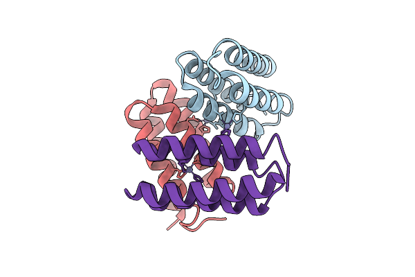 |
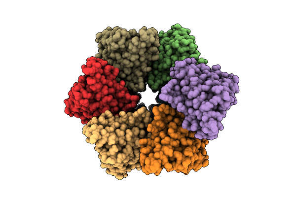
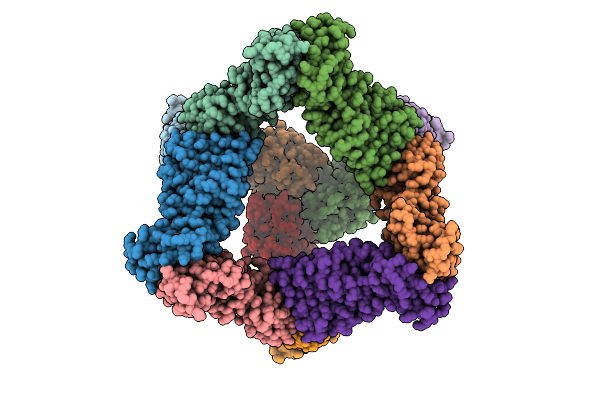
Cryo-Em Structure Of C3 Symmetric Interface Of Designed Zinc-Induced Tetrahedron Cage-T32-Zn1-Hehe-34
Organism: Escherichia coli
Method: ELECTRON MICROSCOPY Release Date: 2025-10-29 Classification: METAL BINDING PROTEIN Ligands: ZN |
 |
Cryo-Em Structure Of Designed Zinc-Induced Tetrahedron Cage-T32-Zn1-Hehe-35
Organism: Escherichia coli
Method: ELECTRON MICROSCOPY Release Date: 2025-10-29 Classification: METAL BINDING PROTEIN Ligands: ZN |