
Search Count: 18
 |
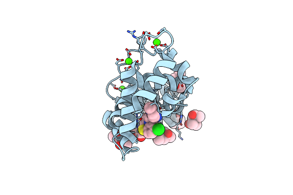
Organism: Homo sapiens
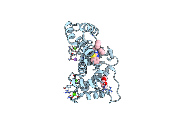
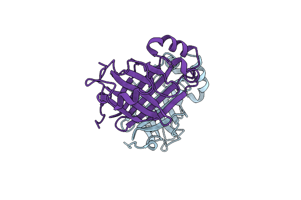
Method: X-RAY DIFFRACTION Release Date: 2025-11-19 Classification: METAL BINDING PROTEIN Ligands: CA, A1IPH, DMS |
 |
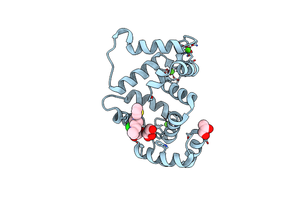
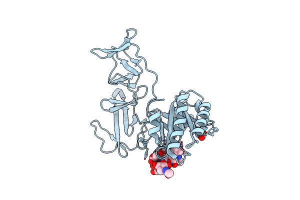
Organism: Homo sapiens
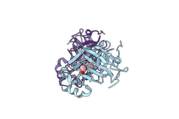
Method: X-RAY DIFFRACTION Release Date: 2025-11-19 Classification: METAL BINDING PROTEIN Ligands: CA, 117, NA, MLI, ACY, SIN, FMT |
 |
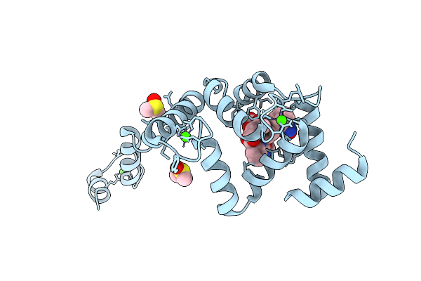
Organism: Homo sapiens
Method: X-RAY DIFFRACTION Release Date: 2025-11-19 Classification: METAL BINDING PROTEIN Ligands: CA, YG7, 1PE, GOL, NA |
 |
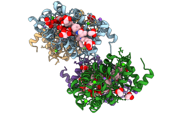
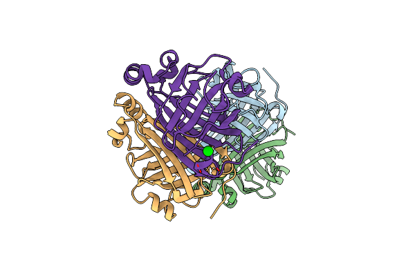
Structure Of Dncs-1 Bound To The Ncs-1/Ric8A Protein/Protein Interaction Regulator Igs-1.76
Organism: Drosophila melanogaster
Method: X-RAY DIFFRACTION Resolution:1.82 Å Release Date: 2018-08-29 Classification: SIGNALING PROTEIN Ligands: CA, BNQ, PG0, NA |
 |
Organism: Drosophila melanogaster
Method: X-RAY DIFFRACTION Resolution:1.60 Å Release Date: 2017-01-25 Classification: CALCIUM-BINDING PROTEIN Ligands: CA, XOC, PG0 |
 |
Organism: Drosophila melanogaster
Method: X-RAY DIFFRACTION Resolution:1.80 Å Release Date: 2017-01-25 Classification: CALCIUM-BINDING PROTEIN Ligands: CA, MPD, FD6 |
 |
Organism: Drosophila melanogaster
Method: X-RAY DIFFRACTION Resolution:1.52 Å Release Date: 2017-01-25 Classification: SIGNALING PROTEIN Ligands: CA, Z80, GOL, EDO |
 |
Crystal Structure Of Single Point Mutant Arg48Gln Of P-Coumaric Acid Decarboxylase From Lactobacillus Plantarum Structural Insights Into The Active Site And Decarboxylation Catalytic Mechanism
Organism: Lactobacillus plantarum
Method: X-RAY DIFFRACTION Resolution:1.73 Å Release Date: 2010-02-09 Classification: LYASE Ligands: BA |
 |
Crystal Structure Of Single Point Mutant Glu71Ser P-Coumaric Acid Decarboxylase
Organism: Lactobacillus plantarum
Method: X-RAY DIFFRACTION Resolution:2.24 Å Release Date: 2010-02-09 Classification: LYASE Ligands: BA, IPA, NA, CL |
 |
Crystal Structure Of P-Coumaric Acid Decarboxylase From Lactobacillus Plantarum: Structural Insights Into The Active Site And Decarboxylation Catalytic Mechanism
Organism: Lactobacillus plantarum
Method: X-RAY DIFFRACTION Resolution:1.38 Å Release Date: 2009-11-17 Classification: LYASE |
 |
Crystal Structure Of Single Point Mutant Tyr20Phe P-Coumaric Acid Decarboxylase From Lactobacillus Plantarum: Structural Insights Into The Active Site And Decarboxylation Catalytic Mechanism
Organism: Lactobacillus plantarum
Method: X-RAY DIFFRACTION Resolution:1.40 Å Release Date: 2009-11-17 Classification: LYASE Ligands: ACT, IPA |
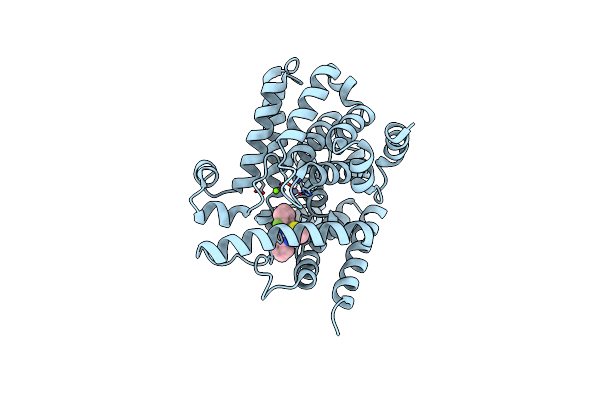 |
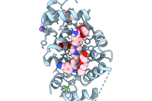
Pde7A Catalytic Domain In Complex With 3-(2,6-Difluorophenyl)-2-(Methylthio)Quinazolin-4(3H)-One
Organism: Homo sapiens
Method: X-RAY DIFFRACTION Resolution:2.40 Å Release Date: 2009-04-28 Classification: HYDROLASE Ligands: TC8, ZN, MG |
 |
Crystal Structure Of Choline Binding Protein F From Streptococcus Pneumoniae In The Presence Of A Peptidoglycan Analogue (Tetrasaccharide-Pentapeptide)
Organism: Streptococcus pneumoniae
Method: X-RAY DIFFRACTION Resolution:2.45 Å Release Date: 2009-02-03 Classification: CHOLINE-BINDING PROTEIN Ligands: CHT |
 |
Crystal Structure Of Choline Binding Protein F From Streptococcus Pneumoniae
Organism: Streptococcus pneumoniae
Method: X-RAY DIFFRACTION Resolution:2.10 Å Release Date: 2008-06-10 Classification: LIPID BINDING PROTEIN Ligands: CHT |
 |
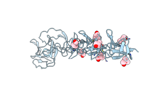
Crystal Structure Of The Modular Cpl-1 Endolysin Complexed With A Peptidoglycan Analogue (Wild-Type Endolysin)
Organism: Streptococcus phage cp-1
Method: X-RAY DIFFRACTION Resolution:2.28 Å Release Date: 2007-07-03 Classification: HYDROLASE Ligands: FMT, ALA, ZGL |
 |
Crystal Structure Of The Modular Cpl-1 Endolysin Complexed With A Peptidoglycan Analogue (E94Q Mutant)
Organism: Streptococcus phage cp-1
Method: X-RAY DIFFRACTION Resolution:1.96 Å Release Date: 2007-07-03 Classification: HYDROLASE Ligands: FMT, ALA, DGN |
 |
Crystal Structure Of The Modular Cpl-1 Endolysin Complexed With A Peptidoglycan Analogue (E94Q Mutant In Complex With A Disaccharide- Pentapeptide)
Organism: Bacteriophage cp-1
Method: X-RAY DIFFRACTION Resolution:1.84 Å Release Date: 2007-07-03 Classification: HYDROLASE Ligands: ALA, DGL, FMT |
 |
Crystal Structure Of The Modular Cpl-1 Endolysin Complexed With A Peptidoglycan Analogue (E94Q Mutant In Complex With A Tetrasaccharide- Pentapeptide)
Organism: Bacteriophage cp-1
Method: X-RAY DIFFRACTION Resolution:1.69 Å Release Date: 2007-07-03 Classification: HYDROLASE Ligands: ALA, DGL, LYS, FMT |