
Search Count: 322
 |
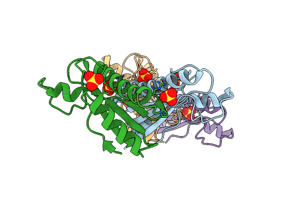
Organism: Porphyromonas gingivalis atcc 33277
Method: ELECTRON MICROSCOPY Release Date: 2025-08-06 Classification: MEMBRANE PROTEIN |
 |
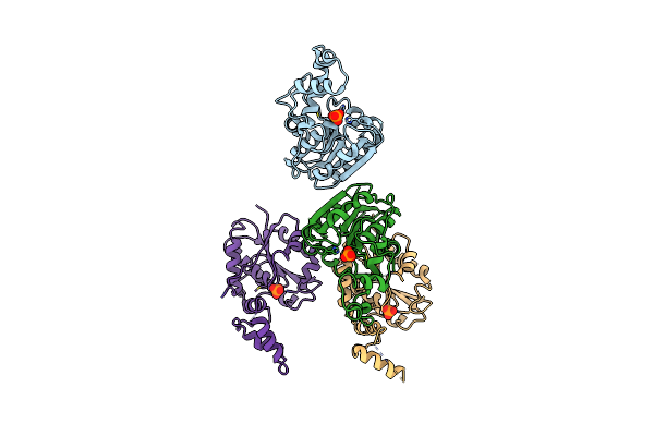
Structure Of The Adherent-Invasive Escherichia Coli Tle3/Tli3 T6Ss Effector/Immunity Complex
Organism: Escherichia coli
Method: X-RAY DIFFRACTION Resolution:3.60 Å Release Date: 2023-02-08 Classification: HYDROLASE Ligands: CA |
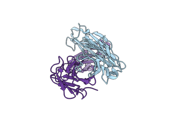 |
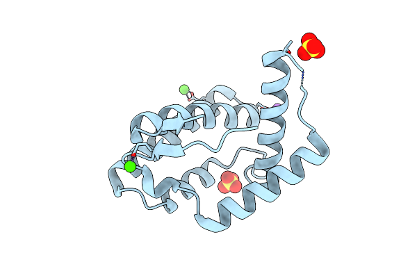
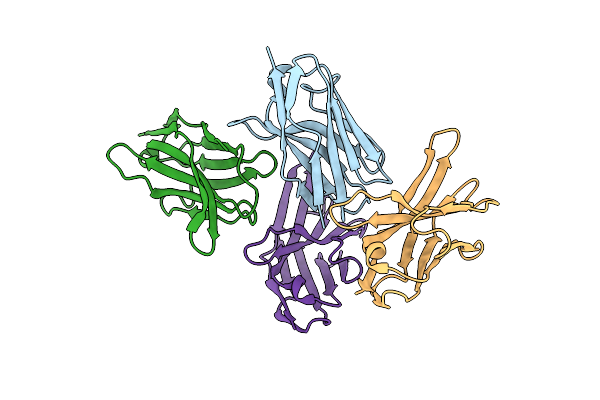
Highly Potent Il6 Antagonistic Antibody Selected From A Camelid Immune Phage Display Repertoire
Organism: Lama glama
Method: X-RAY DIFFRACTION Resolution:2.77 Å Release Date: 2022-09-07 Classification: IMMUNE SYSTEM |
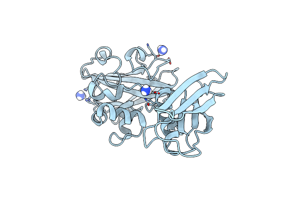 |
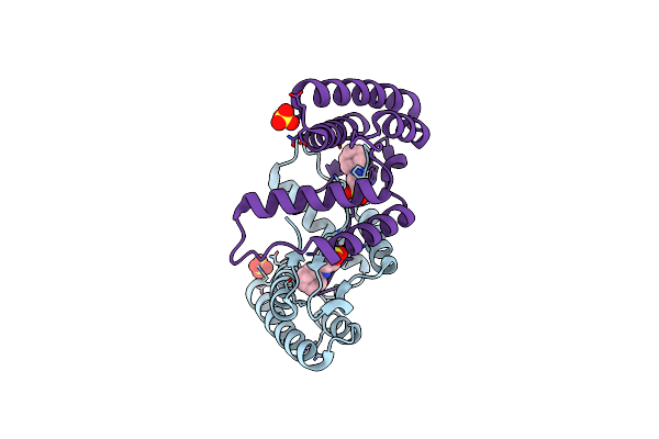
Crystal Structure Of Pseudomonas Aeruginosa Lytic Polysaccharide Monooxygenase Cbpd
Organism: Pseudomonas aeruginosa (strain atcc 15692 / dsm 22644 / cip 104116 / jcm 14847 / lmg 12228 / 1c / prs 101 / pao1)
Method: X-RAY DIFFRACTION Resolution:3.00 Å Release Date: 2022-07-20 Classification: OXIDOREDUCTASE Ligands: NH4 |
 |
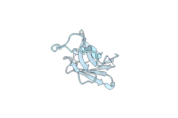
Organism: Escherichia coli
Method: X-RAY DIFFRACTION Resolution:2.43 Å Release Date: 2021-08-04 Classification: PROTEIN BINDING Ligands: SO4 |
 |
Organism: Varroa destructor
Method: X-RAY DIFFRACTION Resolution:1.81 Å Release Date: 2021-07-07 Classification: TRANSPORT PROTEIN Ligands: SO4, CA, NA |
 |
Organism: Varroa destructor
Method: X-RAY DIFFRACTION Resolution:1.20 Å Release Date: 2021-07-07 Classification: TRANSPORT PROTEIN Ligands: SO4, NHE, NA |
 |
Organism: Lama glama
Method: X-RAY DIFFRACTION Resolution:1.70 Å Release Date: 2021-05-26 Classification: IMMUNE SYSTEM |
 |
Organism: Lama glama
Method: X-RAY DIFFRACTION Resolution:2.59 Å Release Date: 2021-05-26 Classification: IMMUNE SYSTEM |
 |
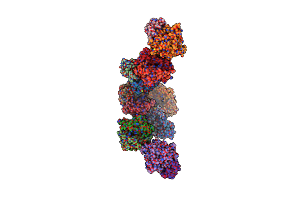
Topological Model Of The P2 Virion Baseplate In Activated Conformation (Closed Tal Trimer)
Organism: Lactococcus phage p2
Method: ELECTRON MICROSCOPY Release Date: 2020-08-26 Classification: VIRAL PROTEIN |
 |
Organism: Lactococcus phage p2
Method: ELECTRON MICROSCOPY Release Date: 2020-08-26 Classification: VIRAL PROTEIN |
 |
Organism: Lactococcus phage p2
Method: ELECTRON MICROSCOPY Release Date: 2020-08-26 Classification: VIRAL PROTEIN |
 |
Structure Of The Pore C-Terminal Domain, A Protein Of T9Ss From Porphyromonas Gingivalis
Organism: Porphyromonas gingivalis jcvi sc001
Method: X-RAY DIFFRACTION Resolution:1.55 Å Release Date: 2020-06-17 Classification: PROTEIN BINDING Ligands: MLD, NA |
 |
Organism: Streptococcus phage d1811, Streptococcus thermophilus (strain atcc baa-491 / lmd-9), Streptococcus thermophilus, Streptococcus virus 2972
Method: ELECTRON MICROSCOPY Resolution:3.20 Å Release Date: 2019-10-02 Classification: HYDROLASE |
 |
Organism: Streptococcus phage d1811, Streptococcus thermophilus (strain atcc baa-491 / lmd-9), Streptococcus thermophilus, Streptococcus virus 2972
Method: ELECTRON MICROSCOPY Resolution:3.00 Å Release Date: 2019-10-02 Classification: HYDROLASE |
 |
Organism: Streptococcus thermophilus dgcc 7710, Streptococcus phage d1811, Streptococcus phage 2972
Method: ELECTRON MICROSCOPY Release Date: 2019-10-02 Classification: HYDROLASE |
 |
Organism: Streptococcus phage d1811, Streptococcus thermophilus dgcc 7710, Streptococcus thermophilus, Streptococcus virus 2972
Method: ELECTRON MICROSCOPY Release Date: 2019-10-02 Classification: HYDROLASE |
 |
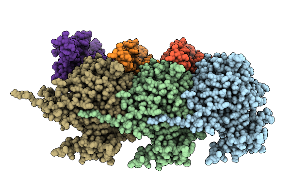
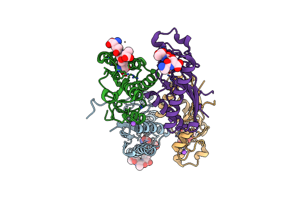
Structure Of The Type Vi Secretion System Tssk-Tssf-Tssg Baseplate Subcomplex Revealed By Cryo-Electron Microscopy - Full Map Sharpened
Organism: Escherichia coli o44:h18 (strain 042 / eaec)
Method: ELECTRON MICROSCOPY Release Date: 2018-12-26 Classification: PROTEIN TRANSPORT |
 |
Organism: Mycobacterium tuberculosis
Method: X-RAY DIFFRACTION Resolution:2.60 Å Release Date: 2018-10-24 Classification: HYDROLASE Ligands: E9H, MPD, CA |
 |
Organism: Mycobacterium tuberculosis cdc1551
Method: X-RAY DIFFRACTION Resolution:2.75 Å Release Date: 2018-10-24 Classification: HYDROLASE Ligands: PO4 |