
Search Count: 21
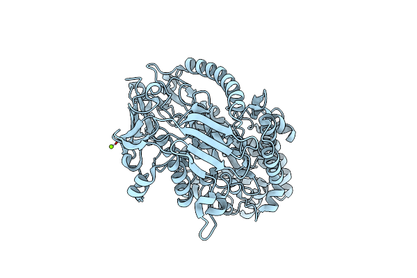 |
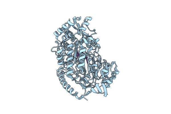
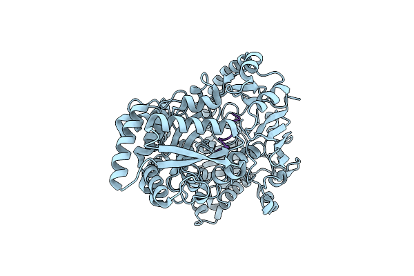
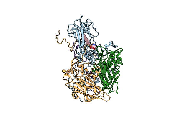
Crystal Structure Of The Pneumococcal Substrate-Binding Protein Alid In Open Conformation
Organism: Streptococcus pneumoniae
Method: X-RAY DIFFRACTION Resolution:1.80 Å Release Date: 2024-05-22 Classification: PEPTIDE BINDING PROTEIN Ligands: MG |
 |
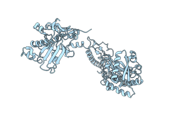
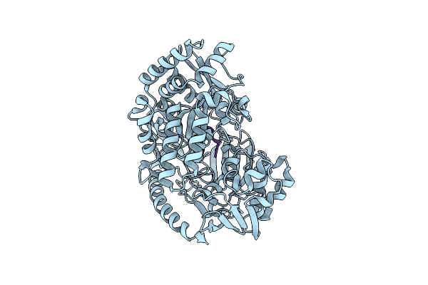
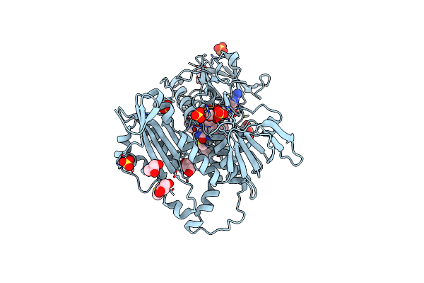
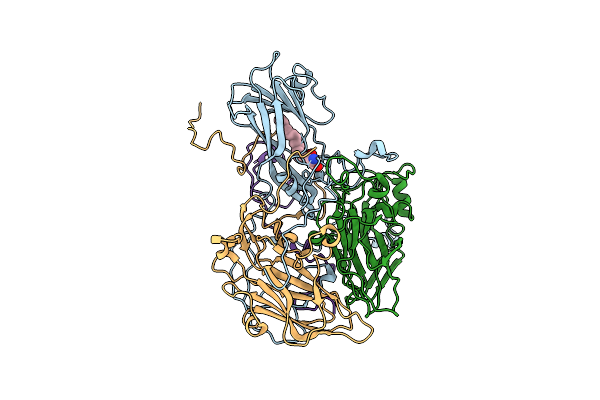
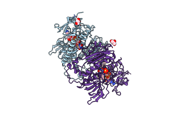
Crystal Structure Of The Pneumococcal Substrate-Binding Protein Alid In Closed Conformation In Complex With Peptide 1
Organism: Streptococcus pneumoniae, Prevotella
Method: X-RAY DIFFRACTION Resolution:2.10 Å Release Date: 2024-05-22 Classification: PEPTIDE BINDING PROTEIN Ligands: ZN |
 |
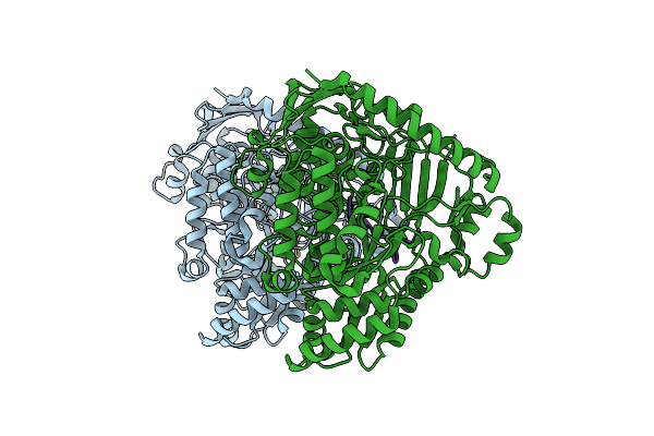
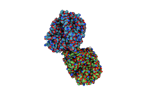
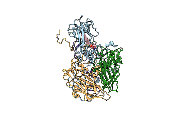
Crystal Structure Of The Pneumococcal Substrate-Binding Protein Alic As A Domain-Swapped Dimer
Organism: Streptococcus pneumoniae
Method: X-RAY DIFFRACTION Resolution:2.38 Å Release Date: 2024-05-22 Classification: PEPTIDE BINDING PROTEIN |
 |
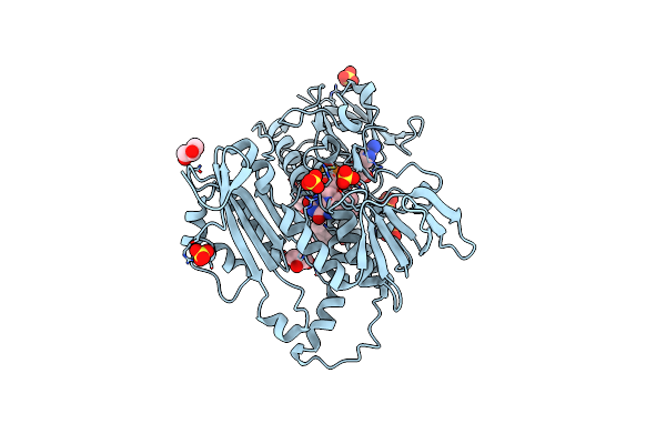
Crystal Structure Of The Pneumococcal Substrate-Binding Protein Alib In Complex With An Unknown Peptide
Organism: Streptococcus pneumoniae, Escherichia coli bl21(de3)
Method: X-RAY DIFFRACTION Resolution:1.65 Å Release Date: 2024-05-22 Classification: PEPTIDE BINDING PROTEIN |
 |
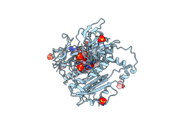
Crystal Structure Of The Pneumococcal Substrate-Binding Protein Alib In Complex With Peptide 2
Organism: Streptococcus pneumoniae, Synthetic construct
Method: X-RAY DIFFRACTION Resolution:2.29 Å Release Date: 2024-05-22 Classification: PEPTIDE BINDING PROTEIN |
 |
Crystal Structure Of The Pneumococcal Substrate-Binding Protein Alib In Complex With Peptide 3
Organism: Streptococcus pneumoniae, Synthetic construct
Method: X-RAY DIFFRACTION Resolution:1.66 Å Release Date: 2024-05-22 Classification: PEPTIDE BINDING PROTEIN |
 |
Crystal Structure Of The Pneumococcal Substrate-Binding Protein Alib In Complex With Peptide 4
Organism: Streptococcus pneumoniae, Synthetic construct
Method: X-RAY DIFFRACTION Resolution:1.49 Å Release Date: 2024-05-22 Classification: PEPTIDE BINDING PROTEIN |
 |
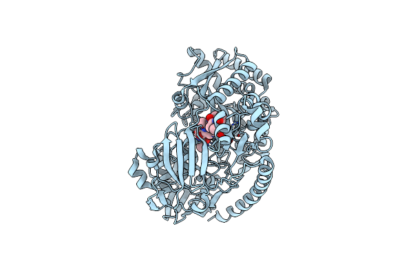
Crystal Structure Of The Pneumococcal Substrate-Binding Protein Amia In Complex With Peptide 5
Organism: Streptococcus pneumoniae, Bacteria
Method: X-RAY DIFFRACTION Resolution:1.76 Å Release Date: 2024-05-22 Classification: PEPTIDE BINDING PROTEIN |
 |
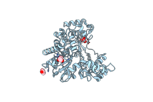
Crystal Structure Of The Pneumococcal Substrate-Binding Protein Amia In Complex With An Unknown Peptide
Organism: Streptococcus pneumoniae, Escherichia coli
Method: X-RAY DIFFRACTION Resolution:1.50 Å Release Date: 2023-12-20 Classification: PEPTIDE BINDING PROTEIN |
 |
Organism: Leishmania infantum
Method: X-RAY DIFFRACTION Resolution:2.50 Å Release Date: 2020-11-18 Classification: FLAVOPROTEIN Ligands: FAD, DMS, GOL, SO4, MWT |
 |
Organism: Leishmania infantum
Method: X-RAY DIFFRACTION Resolution:2.80 Å Release Date: 2020-11-18 Classification: FLAVOPROTEIN Ligands: FAD, SO4, GOL, MWW |
 |
Organism: Leishmania infantum
Method: X-RAY DIFFRACTION Resolution:3.00 Å Release Date: 2020-11-18 Classification: FLAVOPROTEIN Ligands: MWZ, SO4, GOL, FAD, DMS |
 |
Organism: Enterovirus a71
Method: ELECTRON MICROSCOPY Release Date: 2019-12-18 Classification: VIRUS Ligands: SPH |
 |
Organism: Enterovirus a71
Method: ELECTRON MICROSCOPY Release Date: 2019-12-18 Classification: VIRUS Ligands: SPH |
 |
Organism: Enterovirus a71
Method: ELECTRON MICROSCOPY Release Date: 2019-12-18 Classification: VIRUS Ligands: SPH |
 |
Organism: Foot-and-mouth disease virus (isolate -/spain/s8c1santapau/1970 serotype c)
Method: X-RAY DIFFRACTION Resolution:2.30 Å Release Date: 2019-07-03 Classification: VIRAL PROTEIN Ligands: GOL |
 |
Organism: Leishmania infantum
Method: X-RAY DIFFRACTION Resolution:3.30 Å Release Date: 2019-05-15 Classification: FLAVOPROTEIN Ligands: FAD, GOL, SO4, H6H |
 |
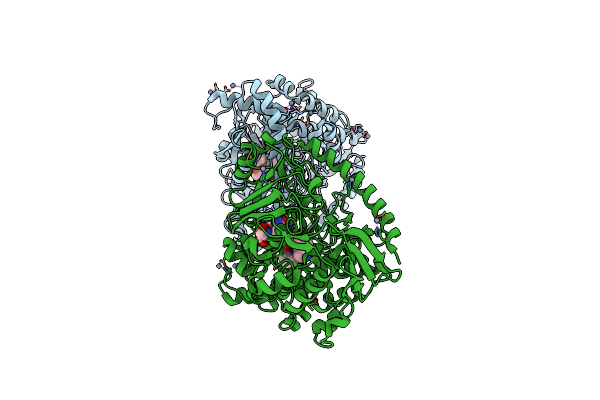
Organism: Rattus norvegicus, Gallus gallus, Bos taurus
Method: X-RAY DIFFRACTION Resolution:2.15 Å Release Date: 2018-03-21 Classification: CELL CYCLE Ligands: GTP, MG, CA, GOL, DLW, GDP, MES, PG4, ACP |
 |
Organism: Rattus norvegicus, Gallus gallus, Bos taurus
Method: X-RAY DIFFRACTION Resolution:2.10 Å Release Date: 2018-03-21 Classification: CELL CYCLE Ligands: GTP, MG, CA, GOL, GDP, MES, DLK, ACP |
 |
Organism: Rattus norvegicus, Gallus gallus, Bos taurus
Method: X-RAY DIFFRACTION Resolution:2.39 Å Release Date: 2016-06-08 Classification: CELL CYCLE Ligands: GTP, MG, CA, GOL, 6NL, GDP, MES, ACP |