Planned Maintenance: Some services may turn out to be unavailable from 15th January, 2026 to 16th January, 2026. We apologize for the inconvenience!

Planned Maintenance: Some services may turn out to be unavailable from 15th January, 2026 to 16th January, 2026. We apologize for the inconvenience!
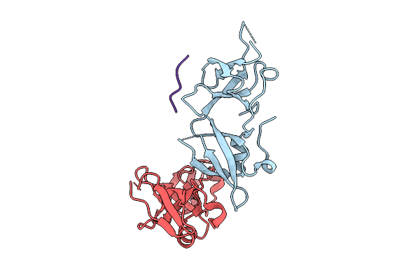 |
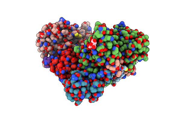
Crystal Structure Of Mouse Bahcc1 Ttd Domain In Complex With H4K20Me1 Peptide
Organism: Mus musculus, Homo sapiens
Method: X-RAY DIFFRACTION Release Date: 2025-07-16 Classification: PROTEIN BINDING |
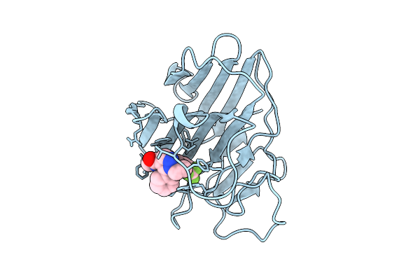 |
Organism: Homo sapiens
Method: X-RAY DIFFRACTION Release Date: 2025-07-16 Classification: LIGASE Ligands: A1EMT |
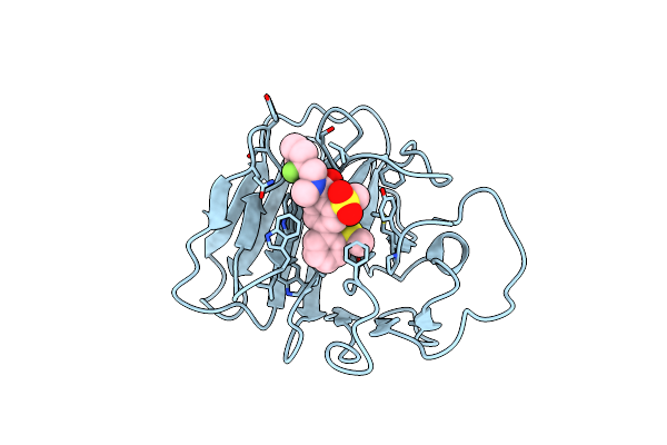 |
Organism: Homo sapiens
Method: X-RAY DIFFRACTION Resolution:1.49 Å Release Date: 2025-05-07 Classification: LIGASE Ligands: A1D9F |
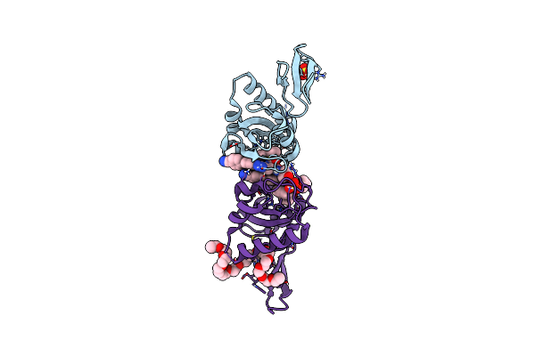 |
Organism: Homo sapiens
Method: X-RAY DIFFRACTION Resolution:1.93 Å Release Date: 2024-09-25 Classification: ISOMERASE Ligands: A1D9K, SO4, PE3 |
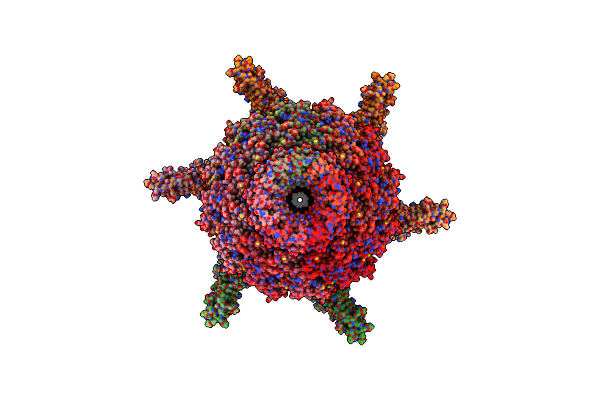 |
Organism: Prochlorococcus phage p-scsp1u
Method: ELECTRON MICROSCOPY Release Date: 2023-11-01 Classification: VIRUS |
 |
Organism: Prochlorococcus phage p-scsp1u
Method: ELECTRON MICROSCOPY Release Date: 2023-10-25 Classification: VIRUS |
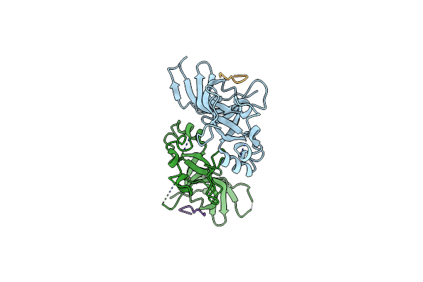 |
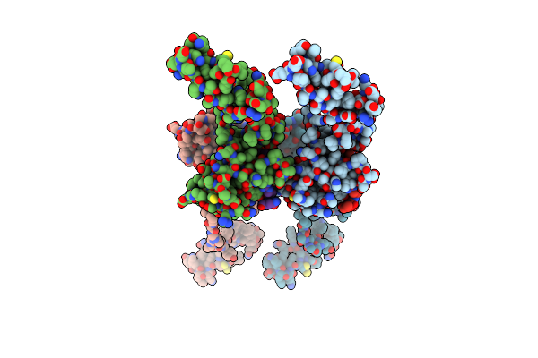
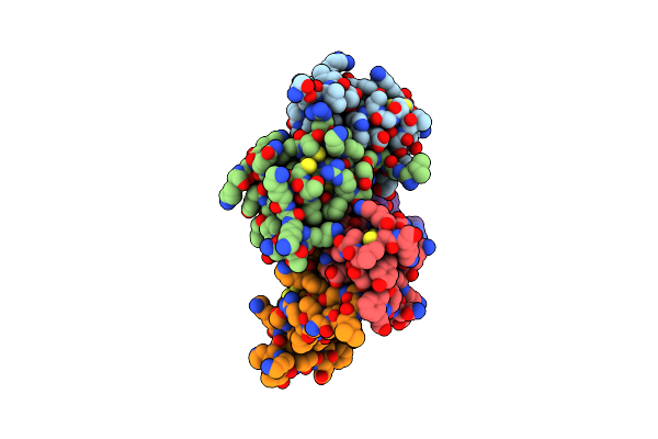
Crystal Structure Of Human Tnrc18 Bah Domain In Complex With H3K9Me3 Peptide
Organism: Homo sapiens
Method: X-RAY DIFFRACTION Resolution:1.84 Å Release Date: 2023-08-02 Classification: STRUCTURAL PROTEIN |
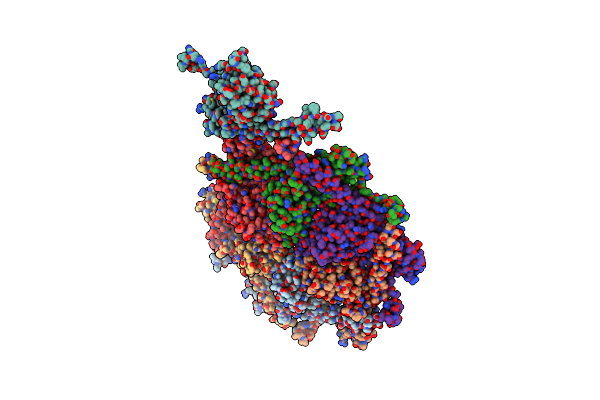 |
Organism: Dinoroseobacter phage vb_dshs-r4c
Method: ELECTRON MICROSCOPY Release Date: 2023-07-12 Classification: VIRAL PROTEIN |
 |
Organism: Dinoroseobacter phage vb_dshs-r4c
Method: ELECTRON MICROSCOPY Release Date: 2023-07-12 Classification: VIRAL PROTEIN |
 |
Organism: Dinoroseobacter phage vb_dshs-r4c
Method: ELECTRON MICROSCOPY Release Date: 2023-07-12 Classification: VIRAL PROTEIN |
 |
Organism: Dinoroseobacter phage vb_dshs-r4c
Method: ELECTRON MICROSCOPY Release Date: 2023-07-12 Classification: VIRUS |
 |
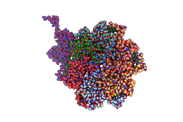
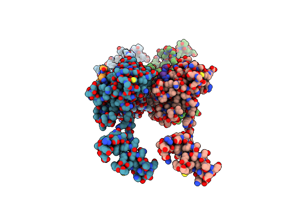
Cryo-Em Model Of The Marine Siphophage Vb_Dshs-R4C Stopper-Terminator Complex
Organism: Dinoroseobacter phage vb_dshs-r4c
Method: ELECTRON MICROSCOPY Release Date: 2023-07-12 Classification: VIRUS |
 |
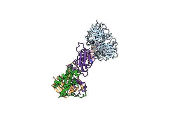
Crystal Structure Of Protac Ms33 In Complex With The Wd Repeat-Containing Protein 5 And Pvhl:Elonginc:Elonginb
Organism: Homo sapiens
Method: X-RAY DIFFRACTION Resolution:1.70 Å Release Date: 2021-10-06 Classification: PROTEIN BINDING Ligands: SCN, EDO, VKA, GOL, NA |
 |
Crystal Structure Of Protac Ms67 In Complex With The Wd Repeat-Containing Protein 5 And Pvhl:Elonginc:Elonginb
Organism: Homo sapiens
Method: X-RAY DIFFRACTION Resolution:2.12 Å Release Date: 2021-10-06 Classification: PROTEIN BINDING Ligands: GOL, X6M |
 |
Organism: Bos taurus, Homo sapiens
Method: X-RAY DIFFRACTION Resolution:2.65 Å Release Date: 2021-02-17 Classification: TRANSFERASE Ligands: ZN |
 |
Organism: Bos taurus, Homo sapiens
Method: X-RAY DIFFRACTION Resolution:2.80 Å Release Date: 2021-02-17 Classification: TRANSFERASE Ligands: ZN |
 |
Organism: Mus musculus, Homo sapiens
Method: X-RAY DIFFRACTION Resolution:3.30 Å Release Date: 2020-09-16 Classification: STRUCTURAL PROTEIN |
 |
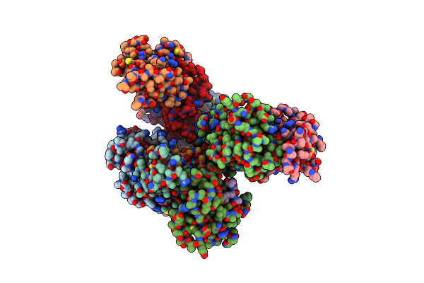
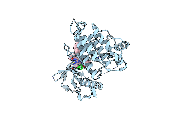
Crystal Structure Of Bovine Dnmt1 Rfts Domain In Complex With H3K9Me3 And Ubiquitin
Organism: Homo sapiens, Bos taurus
Method: X-RAY DIFFRACTION Resolution:3.01 Å Release Date: 2020-07-15 Classification: DNA BINDING PROTEIN Ligands: FLC, ZN |
 |
Organism: Homo sapiens
Method: X-RAY DIFFRACTION Resolution:2.90 Å Release Date: 2020-07-15 Classification: METAL BINDING PROTEIN Ligands: ZN |
 |
Organism: Homo sapiens
Method: X-RAY DIFFRACTION Resolution:1.96 Å Release Date: 2018-12-12 Classification: TRANSFERASE Ligands: 6GY |