
Search Count: 71
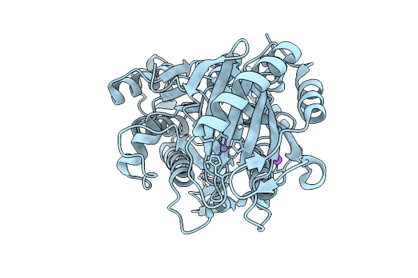 |
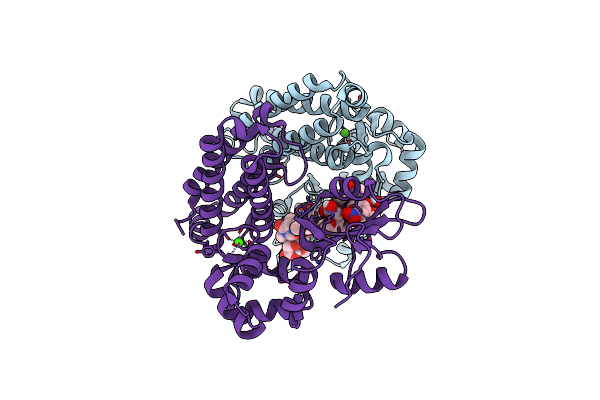
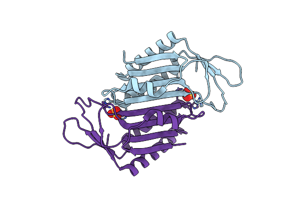
Organism: Citrobacter sp. rhbstw-00271
Method: X-RAY DIFFRACTION Release Date: 2025-09-24 Classification: LIGASE Ligands: ZN, NA |
 |
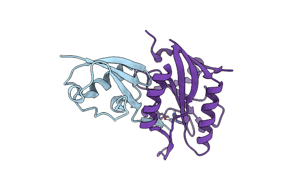
Organism: Citrobacter sp. rhbstw-00271
Method: X-RAY DIFFRACTION Release Date: 2025-09-24 Classification: LIGASE |
 |
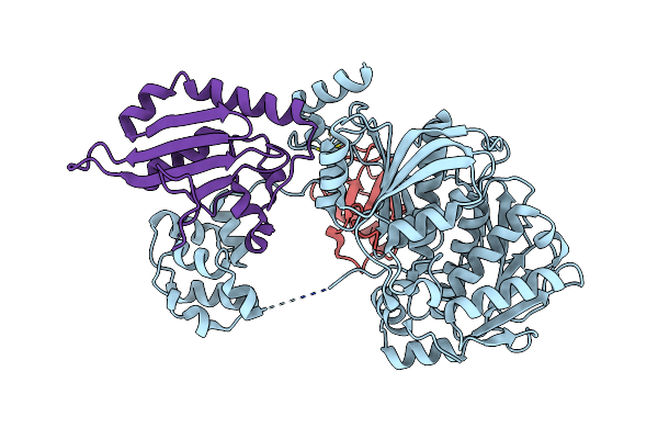
Organism: Citrobacter sp. rhbstw-00271
Method: X-RAY DIFFRACTION Release Date: 2025-09-24 Classification: LIGASE |
 |
Organism: Citrobacter sp. rhbstw-00271
Method: X-RAY DIFFRACTION Release Date: 2025-09-24 Classification: LIGASE |
 |
Organism: Citrobacter sp. rhbstw-00271
Method: X-RAY DIFFRACTION Release Date: 2025-09-24 Classification: LIGASE |
 |
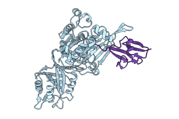
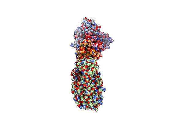
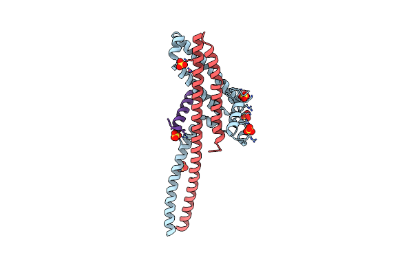
Organism: Severe acute respiratory syndrome coronavirus 2, Enterobacteria phage t4, Synthetic construct, Homo sapiens
Method: ELECTRON MICROSCOPY Release Date: 2025-01-22 Classification: VIRAL PROTEIN |
 |
Organism: Severe acute respiratory syndrome coronavirus 2, Enterobacteria phage t4, Synthetic construct, Homo sapiens
Method: ELECTRON MICROSCOPY Release Date: 2025-01-22 Classification: VIRAL PROTEIN |
 |
Organism: Severe acute respiratory syndrome coronavirus 2, Enterobacteria phage t4, Synthetic construct, Homo sapiens
Method: ELECTRON MICROSCOPY Release Date: 2025-01-22 Classification: VIRAL PROTEIN |
 |
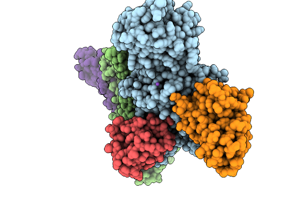
Organism: Severe acute respiratory syndrome coronavirus 2, Tequatrovirus t4, Synthetic construct, Homo sapiens
Method: ELECTRON MICROSCOPY Release Date: 2025-01-22 Classification: VIRAL PROTEIN |
 |
Organism: Homo sapiens
Method: X-RAY DIFFRACTION Resolution:2.40 Å Release Date: 2024-07-03 Classification: TRANSFERASE/TRANSFERASE INHIBITOR Ligands: MG, A1AEG, GOL, PEG |
 |
Organism: Homo sapiens
Method: X-RAY DIFFRACTION Resolution:1.76 Å Release Date: 2024-07-03 Classification: TRANSFERASE/TRANSFERASE INHIBITOR Ligands: MG, CA, A1AD4 |
 |
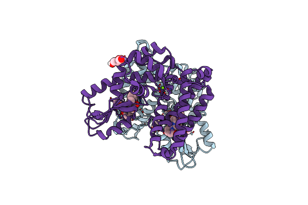
Organism: Ensifer aridi
Method: X-RAY DIFFRACTION Resolution:1.36 Å Release Date: 2024-06-12 Classification: ANTIVIRAL PROTEIN Ligands: ZN |
 |
Organism: Ensifer aridi
Method: X-RAY DIFFRACTION Resolution:1.68 Å Release Date: 2024-06-12 Classification: ANTIVIRAL PROTEIN Ligands: ZN |
 |
Organism: Ensifer aridi
Method: X-RAY DIFFRACTION Resolution:2.68 Å Release Date: 2024-06-12 Classification: ANTIVIRAL PROTEIN Ligands: ZN |
 |
Organism: Ensifer aridi
Method: X-RAY DIFFRACTION Resolution:2.47 Å Release Date: 2024-06-12 Classification: ANTIVIRAL PROTEIN Ligands: ZN |
 |
Organism: Deinococcus radiodurans r1
Method: X-RAY DIFFRACTION Resolution:2.00 Å Release Date: 2023-10-18 Classification: TRANSFERASE Ligands: SO4 |
 |
Organism: Saccharomyces cerevisiae
Method: X-RAY DIFFRACTION Resolution:1.73 Å Release Date: 2023-09-27 Classification: CELL CYCLE Ligands: SO4 |
 |
Organism: Homo sapiens
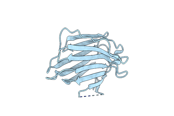
Method: X-RAY DIFFRACTION Resolution:1.60 Å Release Date: 2023-07-05 Classification: SUGAR BINDING PROTEIN |
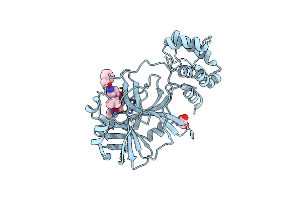 |
Crystal Structure Of The Sars-Cov-2 (Covid-19) Main Protease In Complex With Inhibitor 11
Organism: Severe acute respiratory syndrome coronavirus 2
Method: X-RAY DIFFRACTION Resolution:1.85 Å Release Date: 2023-04-05 Classification: VIRAL PROTEIN/INHIBITOR Ligands: GOL, U6Y |
 |
Crystal Structure Of The Sars-Cov-2 (Covid-19) Main Protease In Complex With Inhibitor 17
Organism: Severe acute respiratory syndrome coronavirus 2
Method: X-RAY DIFFRACTION Resolution:2.20 Å Release Date: 2023-04-05 Classification: VIRAL PROTEIN/INHIBITOR Ligands: U76, GOL |