
Search Count: 19
 |
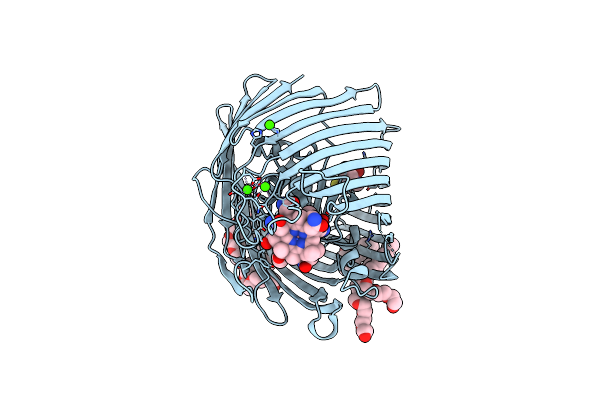
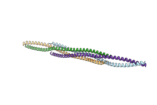
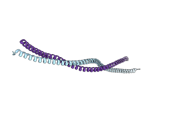
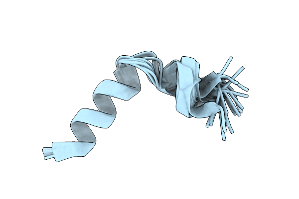
Structure Of The Ebola Virus Envelope Protein Mper/Tm Domain And Its Interaction With The Fusion Loop Explains Their Fusion Activity
Organism: Zaire ebolavirus (strain kikwit-95)
Method: SOLUTION NMR Release Date: 2017-08-30 Classification: VIRAL PROTEIN |
 |
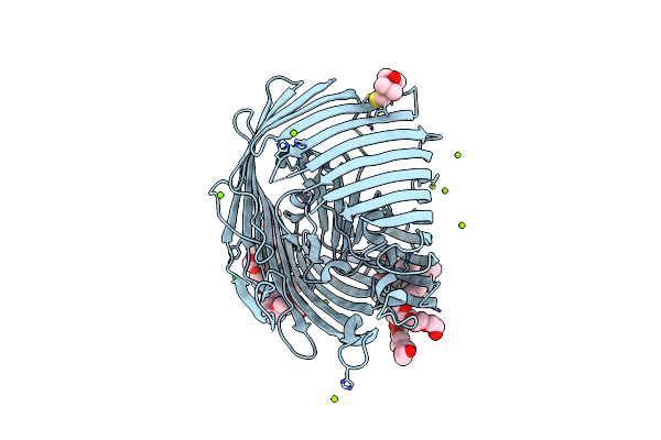
Organism: Mus musculus
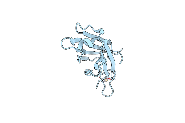
Method: X-RAY DIFFRACTION Resolution:1.95 Å Release Date: 2017-06-14 Classification: METAL BINDING PROTEIN Ligands: CD |
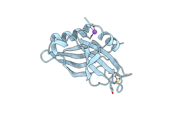 |
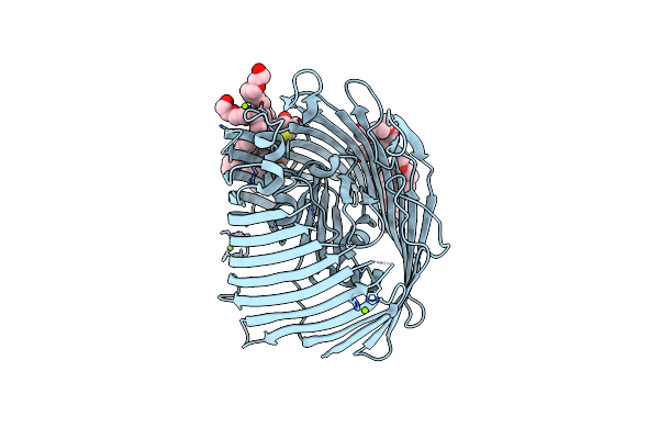
Organism: Mus musculus
Method: X-RAY DIFFRACTION Resolution:1.42 Å Release Date: 2017-06-14 Classification: METAL BINDING PROTEIN Ligands: CD, NA |
 |
Organism: Homo sapiens
Method: X-RAY DIFFRACTION Resolution:1.70 Å Release Date: 2013-07-31 Classification: DNA BINDING PROTEIN |
 |
Organism: Homo sapiens
Method: X-RAY DIFFRACTION Resolution:2.10 Å Release Date: 2013-07-31 Classification: DNA BINDING PROTEIN |
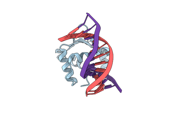 |
Organism: Homo sapiens
Method: X-RAY DIFFRACTION Resolution:2.77 Å Release Date: 2013-07-31 Classification: DNA BINDING PROTEIN/DNA |
 |
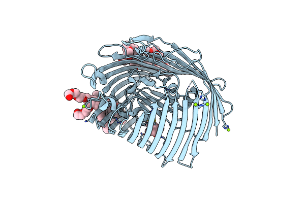
Organism: Escherichia coli
Method: X-RAY DIFFRACTION Resolution:2.60 Å Release Date: 2011-10-26 Classification: TRANSPORT PROTEIN Ligands: C8E, MG, MTN |
 |
Organism: Escherichia coli
Method: X-RAY DIFFRACTION Resolution:2.30 Å Release Date: 2011-10-26 Classification: TRANSPORT PROTEIN Ligands: C8E, MG, MTN |
 |
Organism: Escherichia coli
Method: X-RAY DIFFRACTION Resolution:2.44 Å Release Date: 2010-09-15 Classification: TRANSPORT PROTEIN Ligands: C8E, MTN, MG |
 |
Crystal Structure Of Spin-Labeled Btub V10R1 With Bound Calcium And Cyanocobalamin
Organism: Escherichia coli
Method: X-RAY DIFFRACTION Resolution:2.44 Å Release Date: 2010-09-15 Classification: TRANSPORT PROTEIN Ligands: CNC, C8E, MTN, CA |
 |
Organism: Rattus norvegicus
Method: SOLUTION NMR Release Date: 2009-12-01 Classification: MEMBRANE PROTEIN |
 |
Organism: Escherichia coli
Method: SOLUTION NMR Release Date: 2008-10-07 Classification: MEMBRANE PROTEIN, OXIDOREDUCTASE |
 |
Organism: Escherichia coli
Method: SOLUTION NMR Release Date: 2008-10-07 Classification: MEMBRANE PROTEIN, OXIDOREDUCTASE Ligands: UQ2 |
 |
Organism: Homo sapiens
Method: X-RAY DIFFRACTION Resolution:2.10 Å Release Date: 2007-11-27 Classification: STRUCTURAL PROTEIN Ligands: HG |
 |
Organism: Rattus norvegicus
Method: X-RAY DIFFRACTION Resolution:2.24 Å Release Date: 2007-11-27 Classification: NUCLEAR PROTEIN |
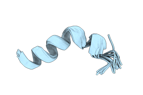 |
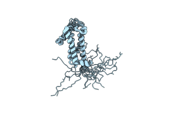
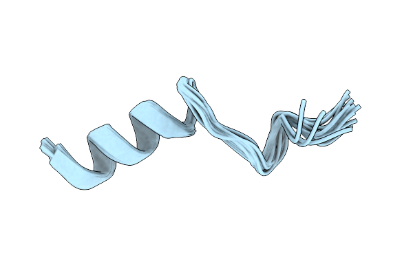
Nmr Structure Of G1S Mutant Of Influenza Hemagglutinin Fusion Peptide In Dpc Micelles At Ph 5
|
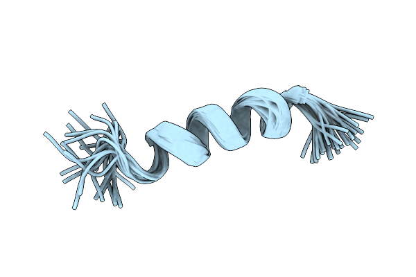 |
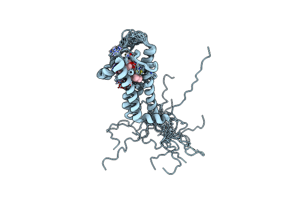
Nmr Structure Of G1V Mutant Of Influenza Hemagglutinin Fusion Peptide In Dpc Micelles At Ph 5
|
 |
 |