
Search Count: 22
 |
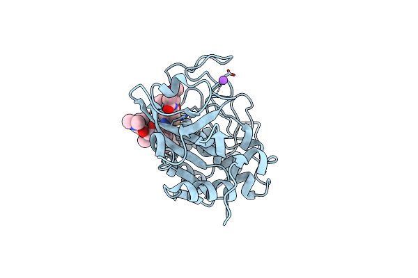
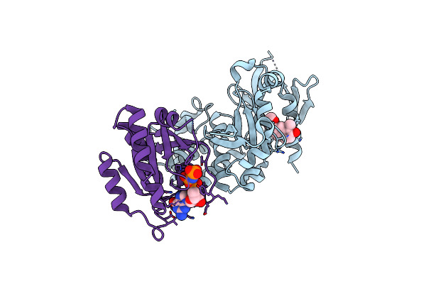
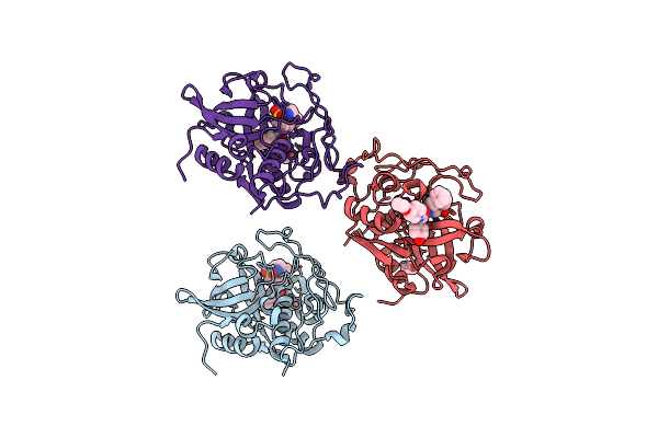
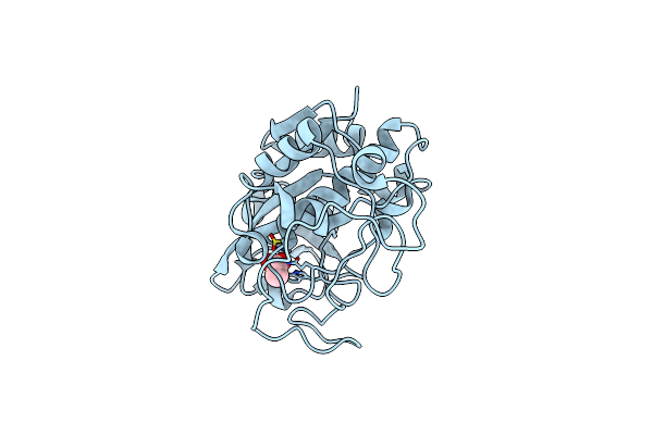
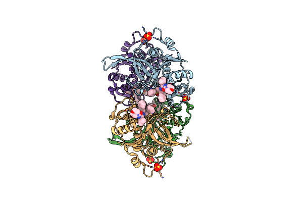
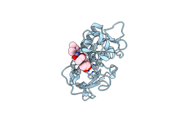
Structure Of Cathepsin B1 From Schistosoma Mansoni (Smcb1) In Complex With A Carborane Inhibitor
Organism: Schistosoma mansoni
Method: X-RAY DIFFRACTION Release Date: 2025-10-29 Classification: HYDROLASE Ligands: A1IHQ, EDO |
 |
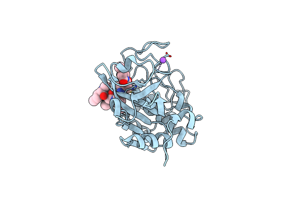
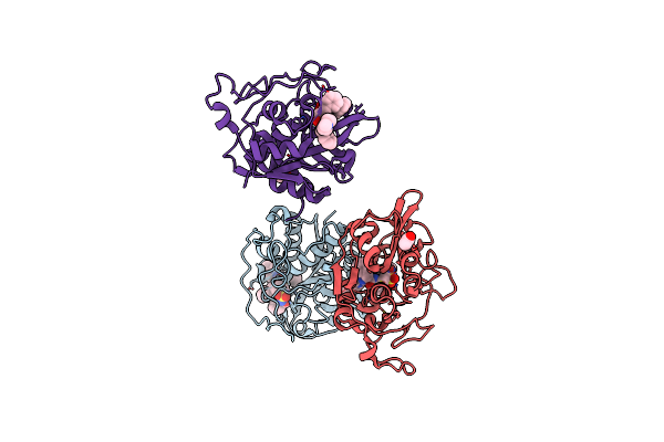
Organism: Schistosoma mansoni
Method: X-RAY DIFFRACTION Resolution:1.55 Å Release Date: 2024-02-21 Classification: HYDROLASE Ligands: UA9, EDO, NA |
 |
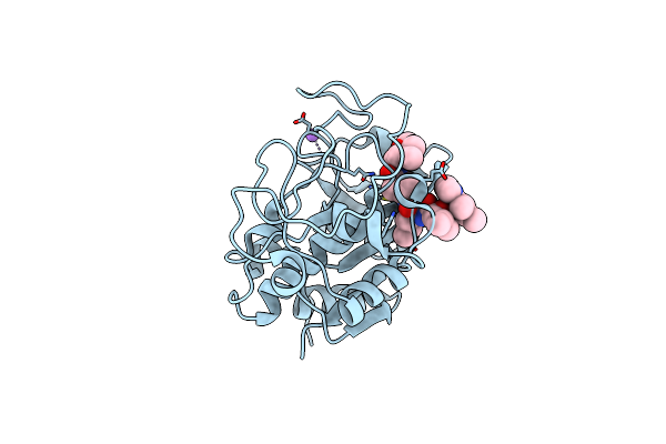
Organism: Schistosoma mansoni
Method: X-RAY DIFFRACTION Resolution:1.20 Å Release Date: 2024-02-07 Classification: HYDROLASE Ligands: GN9, NA |
 |
Organism: Schistosoma mansoni
Method: X-RAY DIFFRACTION Resolution:1.60 Å Release Date: 2024-02-07 Classification: HYDROLASE Ligands: U9X, NA |
 |
Co-Crystal Structure Of Krit1 With A 1-Hydroxy 2-Naphthaldehyde Derivative (6-Ethynyl-2-Hydroxy-1-Naphthaldehyde)
Organism: Homo sapiens
Method: X-RAY DIFFRACTION Resolution:2.15 Å Release Date: 2023-12-13 Classification: CELL ADHESION Ligands: XHZ, MG, GNP |
 |
Co-Crystal Structure Of Krit1 With A 1-Hydroxy 2-Naphthaldehyde Derivative (6-(Furan-2-Yl)-2-Hydroxy-1-Naphthaldehyde).
Organism: Homo sapiens
Method: X-RAY DIFFRACTION Resolution:2.01 Å Release Date: 2023-12-06 Classification: CELL ADHESION Ligands: XE2, MG, GNP |
 |
Co-Crystal Structure Of Krit1 With A 1-Hydroxy 2-Naphthaldehyde Derivative (6-(Furan-2-Yl)-2-Hydroxy-1-Naphthaldehyde)
Organism: Homo sapiens
Method: X-RAY DIFFRACTION Resolution:2.25 Å Release Date: 2023-12-06 Classification: CELL ADHESION Ligands: ZTA, MG, GNP |
 |
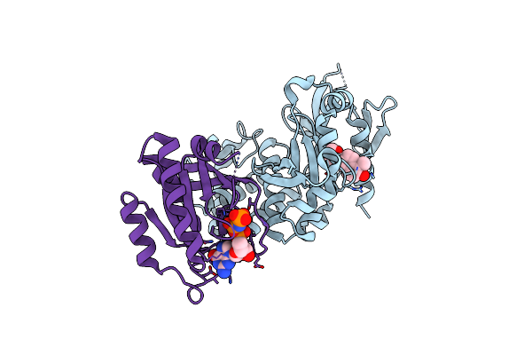
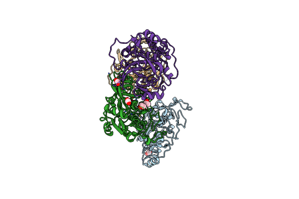
Crystal Structure Of The Schistosoma Mansoni Vkr2 Kinase Domain In An Active-Like State (Adp-Bound)
Organism: Schistosoma mansoni
Method: X-RAY DIFFRACTION Resolution:3.07 Å Release Date: 2022-10-12 Classification: TRANSFERASE Ligands: ADP |
 |
Structure Of Cathepsin B1 From Schistosoma Mansoni (Smcb1) In Complex With An Azanitrile Inhibitor
Organism: Schistosoma mansoni
Method: X-RAY DIFFRACTION Resolution:1.29 Å Release Date: 2020-12-16 Classification: HYDROLASE Ligands: ACT, ORW |
 |
Organism: Fasciola hepatica
Method: X-RAY DIFFRACTION Resolution:1.90 Å Release Date: 2020-02-12 Classification: ISOMERASE Ligands: SO4 |
 |
Structure Of Cathepsin B1 From Schistosoma Mansoni In Complex With Wrr391 Inhibitor
Organism: Schistosoma mansoni
Method: X-RAY DIFFRACTION Resolution:1.91 Å Release Date: 2018-11-21 Classification: HYDROLASE Ligands: 9U8, ACT |
 |
Structure Of Cathepsin B1 From Schistosoma Mansoni In Complex With Wrr286 Inhibitor
Organism: Schistosoma mansoni
Method: X-RAY DIFFRACTION Resolution:1.55 Å Release Date: 2018-11-21 Classification: HYDROLASE Ligands: VS4, ACT |
 |
Organism: Schistosoma mansoni
Method: X-RAY DIFFRACTION Resolution:1.95 Å Release Date: 2014-02-05 Classification: HYDROLASE Ligands: EDO |
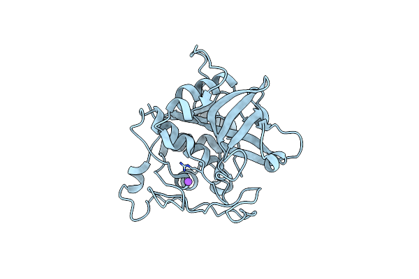 |
Structure Of Intermediate Processing Form Of Cathepsin B1 From Schistosoma Mansoni
Organism: Schistosoma mansoni
Method: X-RAY DIFFRACTION Resolution:1.90 Å Release Date: 2014-02-05 Classification: HYDROLASE |
 |
Organism: Schistosoma mansoni
Method: X-RAY DIFFRACTION Resolution:1.30 Å Release Date: 2014-02-05 Classification: HYDROLASE Ligands: CL, ACT |
 |
Structure Of Cathepsin B1 From Schistosoma Mansoni In Complex With Ca074 Inhibitor
Organism: Schistosoma mansoni
Method: X-RAY DIFFRACTION Resolution:1.30 Å Release Date: 2011-08-10 Classification: HYDROLASE/HYDROLASE INHIBITOR Ligands: 074, PEG, ACT |
 |
Structure Of Cathepsin B1 From Schistosoma Mansoni In Complex With K11017 Inhibitor
Organism: Schistosoma mansoni
Method: X-RAY DIFFRACTION Resolution:1.80 Å Release Date: 2011-08-10 Classification: HYDROLASE/HYDROLASE INHIBITOR Ligands: C1P, ACT |
 |
Structure Of Cathepsin B1 From Schistosoma Mansoni In Complex With K11777 Inhibitor
Organism: Schistosoma mansoni
Method: X-RAY DIFFRACTION Resolution:2.64 Å Release Date: 2011-08-10 Classification: HYDROLASE/HYDROLASE INHIBITOR Ligands: 0IW |
 |
Organism: Plasmodium falciparum
Method: X-RAY DIFFRACTION Resolution:2.42 Å Release Date: 2009-01-20 Classification: HYDROLASE Ligands: C1P, SO4 |
 |
The Crystal Structure Of Rhodesain, The Major Cysteine Protease Of T. Brucei Rhodesiense, Bound To Inhibitor K777
Organism: Trypanosoma brucei rhodesiense
Method: X-RAY DIFFRACTION Resolution:1.65 Å Release Date: 2008-03-25 Classification: HYDROLASE Ligands: D1R |