
Search Count: 1,201
All
Selected
 |
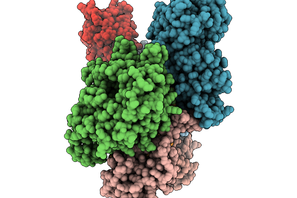
Organism: Plasmodium falciparum 3d7
Method: ELECTRON MICROSCOPY Release Date: 2026-03-11 Classification: STRUCTURAL PROTEIN Ligands: GTP, MG, GDP, TA1 |
 |
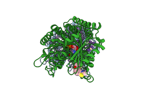
Organism: Plasmodium falciparum 3d7
Method: ELECTRON MICROSCOPY Release Date: 2026-03-11 Classification: STRUCTURAL PROTEIN Ligands: GTP, MG, G2P |
 |
Organism: Homo sapiens, Oryctolagus cuniculus
Method: ELECTRON MICROSCOPY Resolution:3.13 Å Release Date: 2026-03-04 Classification: PROTEIN FIBRIL Ligands: ADP, MG |
 |
Organism: Homo sapiens, Oryctolagus cuniculus
Method: ELECTRON MICROSCOPY Resolution:3.68 Å Release Date: 2026-03-04 Classification: PROTEIN FIBRIL Ligands: ADP, MG |
 |
Organism: Homo sapiens, Oryctolagus cuniculus
Method: ELECTRON MICROSCOPY Resolution:3.50 Å Release Date: 2026-03-04 Classification: PROTEIN FIBRIL Ligands: ADP, MG |
 |
Organism: Homo sapiens, Oryctolagus cuniculus
Method: ELECTRON MICROSCOPY Resolution:3.48 Å Release Date: 2026-03-04 Classification: PROTEIN FIBRIL Ligands: ADP, MG |
 |
Organism: Homo sapiens, Oryctolagus cuniculus
Method: ELECTRON MICROSCOPY Resolution:3.02 Å Release Date: 2026-03-04 Classification: PROTEIN FIBRIL Ligands: ADP, MG |
 |
Organism: Homo sapiens, Oryctolagus cuniculus
Method: ELECTRON MICROSCOPY Resolution:3.18 Å Release Date: 2026-03-04 Classification: PROTEIN FIBRIL Ligands: ADP, MG |
 |
Organism: Homo sapiens, Oryctolagus cuniculus
Method: ELECTRON MICROSCOPY Resolution:3.46 Å Release Date: 2026-03-04 Classification: PROTEIN FIBRIL Ligands: ADP, MG |
 |
Organism: Homo sapiens, Oryctolagus cuniculus
Method: ELECTRON MICROSCOPY Resolution:3.58 Å Release Date: 2026-03-04 Classification: PROTEIN FIBRIL Ligands: ADP, MG |
 |
Organism: Homo sapiens, Oryctolagus cuniculus
Method: ELECTRON MICROSCOPY Resolution:3.06 Å Release Date: 2026-03-04 Classification: PROTEIN FIBRIL Ligands: ADP, MG |
 |
Organism: Homo sapiens, Oryctolagus cuniculus
Method: ELECTRON MICROSCOPY Resolution:3.06 Å Release Date: 2026-03-04 Classification: PROTEIN FIBRIL Ligands: ADP, MG |
 |
Organism: Homo sapiens, Oryctolagus cuniculus
Method: ELECTRON MICROSCOPY Resolution:2.06 Å Release Date: 2026-03-04 Classification: PROTEIN FIBRIL Ligands: ADP, MG |
 |
Organism: Homo sapiens, Oryctolagus cuniculus
Method: ELECTRON MICROSCOPY Resolution:2.62 Å Release Date: 2026-03-04 Classification: PROTEIN FIBRIL Ligands: ADP, MG |
 |
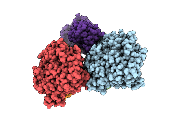
Tubulin Cofactors D,E,G,C And Tubulin Complex -- Tbcc N Terminus Bound To Tubulin
Organism: Saccharomyces cerevisiae, Escherichia coli, Sus scrofa
Method: ELECTRON MICROSCOPY Resolution:3.80 Å Release Date: 2026-02-04 Classification: CYTOSOLIC PROTEIN Ligands: GTP, GDP |
 |
Organism: Homo sapiens
Method: ELECTRON MICROSCOPY Release Date: 2026-01-21 Classification: STRUCTURAL PROTEIN |
 |
Organism: Homo sapiens, Bos taurus
Method: ELECTRON MICROSCOPY Release Date: 2025-11-19 Classification: PROTEIN BINDING Ligands: ATP, MG |
 |
Organism: Mus musculus
Method: ELECTRON MICROSCOPY Resolution:3.19 Å Release Date: 2025-11-12 Classification: STRUCTURAL PROTEIN Ligands: MG, GDP, EPB, GTP |
 |
Organism: Xenopus laevis, Bos taurus
Method: ELECTRON MICROSCOPY Resolution:2.89 Å Release Date: 2025-11-05 Classification: CELL CYCLE Ligands: G2P, MG, GTP |
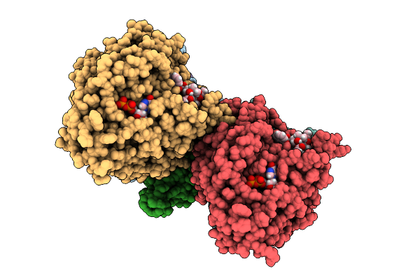 |
Cryo-Em Structure Of Taxol-Microtubules In Complex With The C1 Domain Of Gefh1
Organism: Homo sapiens, Bos taurus
Method: ELECTRON MICROSCOPY Release Date: 2025-10-29 Classification: SIGNALING PROTEIN Ligands: ZN, GTP, MG, GDP, TA1 |