
Search Count: 17
 |
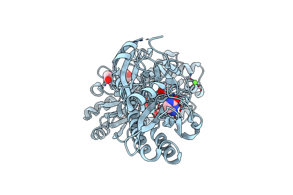
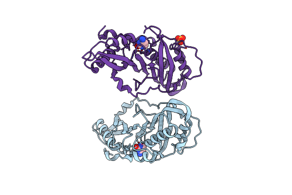
Nucleoside-2'-Deoxyribosyltransferase From Lactobacillus Leichmannii. Complex With Ribose And Cytosine
Organism: Lactobacillus leichmannii
Method: X-RAY DIFFRACTION Resolution:2.41 Å Release Date: 2024-12-25 Classification: TRANSFERASE Ligands: CYT, RIB |
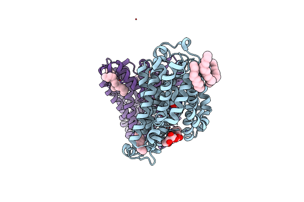 |
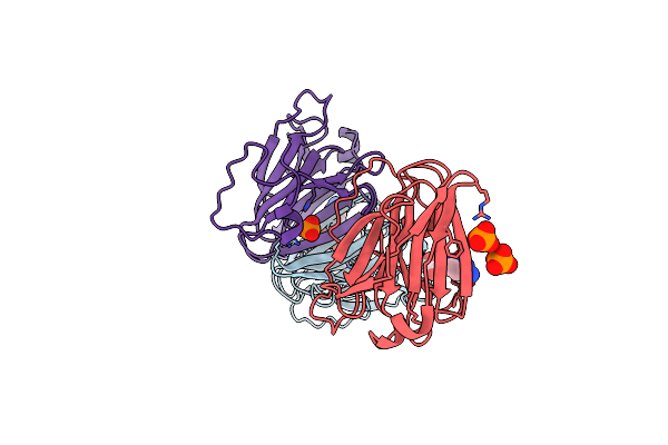
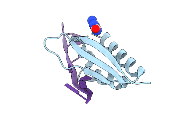
Structure Of Codb, A Cytosine Transporter In An Outward-Facing Conformation
Organism: Proteus vulgaris
Method: X-RAY DIFFRACTION Resolution:2.40 Å Release Date: 2022-07-13 Classification: TRANSPORT PROTEIN Ligands: CYT, PEF, LMT, A6L, NA |
 |
X-Ray Structure Of Uridine Phosphorylase From Vibrio Cholerae In Complex With Cytidine And Cytosine At 1.11 A Resolution
Organism: Vibrio cholerae
Method: X-RAY DIFFRACTION Resolution:1.11 Å Release Date: 2017-08-23 Classification: TRANSFERASE Ligands: EDO, K, CTN, CYT, GOL, SO4, CL |
 |
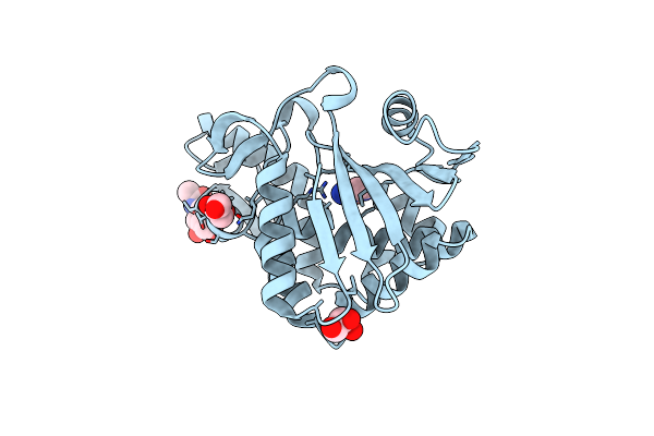
Crystal Structure Of Isoform 2 Of Purine Nucleoside Phosphorylase From Schistosoma Mansoni In Complex With Cytosine
Organism: Schistosoma mansoni
Method: X-RAY DIFFRACTION Resolution:1.36 Å Release Date: 2017-08-09 Classification: TRANSFERASE Ligands: CYT, EDO |
 |
Crystal Structure Of Isoform 2 Of Purine Nucleoside Phosphorylase From Schistosoma Mansoni In Complex With Cytosine And Ribose-1-Phosphate
Organism: Schistosoma mansoni
Method: X-RAY DIFFRACTION Resolution:1.42 Å Release Date: 2017-08-09 Classification: TRANSFERASE Ligands: CYT, R1P |
 |
X-Ray Structure Uridine Phosphorylase From Vibrio Cholerae In Complex With Cytosine At 1.06A.
Organism: Vibrio cholerae
Method: X-RAY DIFFRACTION Resolution:1.06 Å Release Date: 2016-11-23 Classification: TRANSFERASE Ligands: CYT, GOL, MG, NA, CL, TRS, EDO |
 |
Extracellular Ligand Binding Receptor From Desulfohalobium Retbaense Dsm5692
Organism: Desulfohalobium retbaense dsm 5692
Method: X-RAY DIFFRACTION Resolution:2.00 Å Release Date: 2016-08-10 Classification: SIGNALING PROTEIN Ligands: COI, CYT, ACY, CA, GOL, EDO |
 |
Organism: Yersinia enterocolitica subsp. enterocolitica 8081
Method: X-RAY DIFFRACTION Resolution:1.75 Å Release Date: 2015-08-05 Classification: ISOMERASE Ligands: PO4, CYT, EDO |
 |
Structure Of The Complex Of Type 1 Ribosome Inactivating Protein From Momordica Balsamina With A Pyrimidine Base, Cytosine At 1.98 A Resolution
Organism: Momordica balsamina
Method: X-RAY DIFFRACTION Resolution:1.98 Å Release Date: 2015-06-03 Classification: HYDROLASE Ligands: NAG, GOL, CYT |
 |
Organism: Nitrosomonas europaea
Method: X-RAY DIFFRACTION Resolution:3.00 Å Release Date: 2014-01-22 Classification: HYDROLASE Ligands: ZN, CYT |
 |
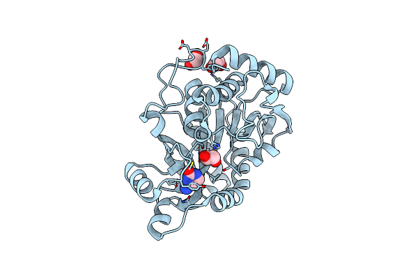
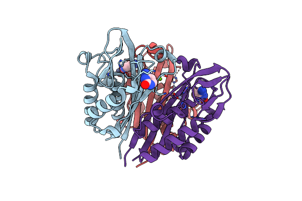
Crystal Structure Of 7-Cyano-7-Deazaguanine Reductase, Quef From Vibrio Cholerae O1 Biovar El Tor Complexed With Cytosine
Organism: Vibrio cholerae o1 biovar el tor
Method: X-RAY DIFFRACTION Resolution:1.50 Å Release Date: 2013-01-23 Classification: OXIDOREDUCTASE Ligands: CYT, CL, SO4 |
 |
Organism: Adeno-associated virus - 1
Method: X-RAY DIFFRACTION Resolution:2.50 Å Release Date: 2011-12-21 Classification: VIRUS Ligands: ADE, CYT |
 |
Organism: Bacillus anthracis
Method: X-RAY DIFFRACTION Resolution:2.00 Å Release Date: 2011-07-06 Classification: TRANSFERASE Ligands: ACO, CYT, MG, EPE, CPS |
 |
Structural Characterization Of The Dual Glycan Binding Adeno-Associated Virus Serotype 6
Organism: Adeno-associated virus - 6
Method: X-RAY DIFFRACTION Resolution:3.00 Å Release Date: 2011-03-23 Classification: VIRUS Ligands: CYT, OAH |
 |
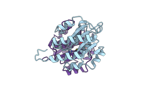
Crystal Structure Of 2C-Methyl-D-Erythritol 2,4-Cyclodiphosphate Synthase From Burkholderia Pseudomallei With Cytosine And Fol Fragment 717, Imidazo[2,1-B][1,3]Thiazol-6-Ylmethanol
Organism: Burkholderia pseudomallei
Method: X-RAY DIFFRACTION Resolution:1.80 Å Release Date: 2010-04-07 Classification: LYASE Ligands: ZN, CYT, 717, TRS |
 |
Crystal Structure Of 2C-Methyl-D-Erythritol 2,4-Cyclodiphosphate Synthase From Burkholderia Pseudomallei With Cytosine
Organism: Burkholderia pseudomallei
Method: X-RAY DIFFRACTION Resolution:2.30 Å Release Date: 2009-08-18 Classification: LYASE Ligands: ZN, CYT, CL, MG |
 |
Crystal Structure Of The Third Kh Domain Of Human Poly(C)-Binding Protein-2 In Complex With C-Rich Strand Of Human Telomeric Dna
Organism: Homo sapiens
Method: X-RAY DIFFRACTION Resolution:1.60 Å Release Date: 2007-06-12 Classification: RNA AND DNA BINDING PROTEIN/DNA Ligands: CYT |