
Search Count: 160
 |
Organism: Gloeobacter violaceus pcc 7421
Method: ELECTRON MICROSCOPY Release Date: 2025-12-24 Classification: PHOTOSYNTHESIS Ligands: CYC |
 |
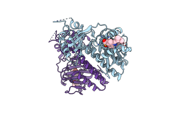
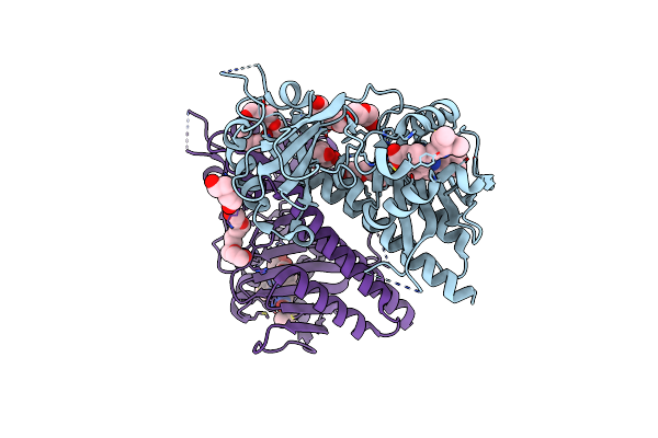
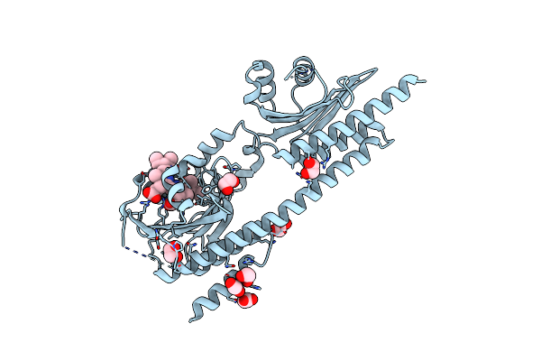
X-Ray Structure Of The Adduct Formed By Dirhodium Tetraacetate With A C-Phycocyanin
Organism: Galdieria phlegrea
Method: X-RAY DIFFRACTION Release Date: 2025-12-17 Classification: PHOTOSYNTHESIS Ligands: CYC, A1JTN, ACT, DOD |
 |
Organism: Gloeobacter violaceus pcc 7421
Method: ELECTRON MICROSCOPY Release Date: 2025-12-03 Classification: PHOTOSYNTHESIS Ligands: CYC |
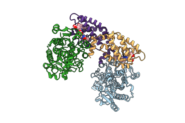 |
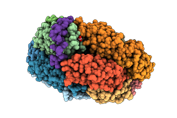
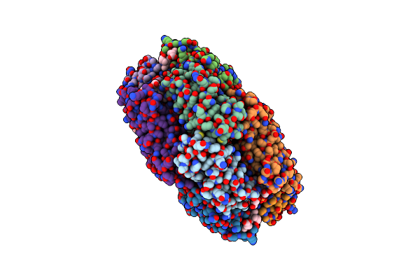
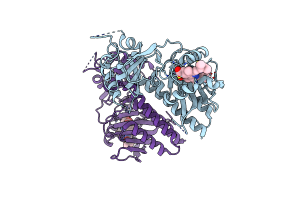
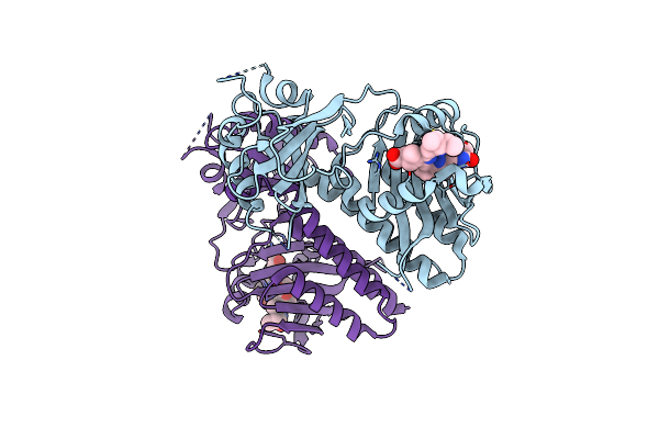
Structure Of The Recombinant Structure Of The Subunit Of Allophycocyanin (Apc) And The Formate Dehydrogenase (Fdh)
Organism: Candida boidinii, Picosynechococcus sp. pcc 7003
Method: X-RAY DIFFRACTION Release Date: 2025-11-05 Classification: PHOTOSYNTHESIS Ligands: CYC |
 |
Organism: Gloeobacter violaceus pcc 7421
Method: ELECTRON MICROSCOPY Release Date: 2025-09-24 Classification: PHOTOSYNTHESIS Ligands: CYC |
 |
Organism: Gloeobacter violaceus pcc 7421
Method: ELECTRON MICROSCOPY Release Date: 2025-09-24 Classification: PHOTOSYNTHESIS Ligands: CYC |
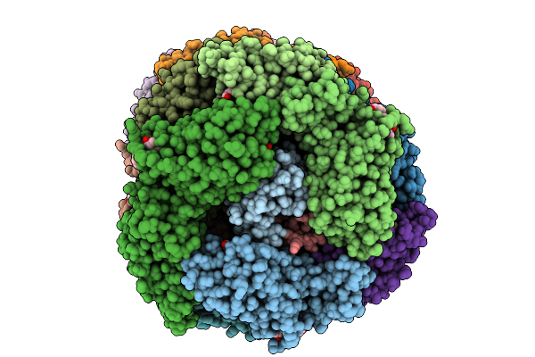 |
Organism: Gloeobacter violaceus pcc 7421
Method: ELECTRON MICROSCOPY Release Date: 2025-09-24 Classification: PHOTOSYNTHESIS Ligands: CYC |
 |
Organism: Gloeobacter violaceus pcc 7421
Method: ELECTRON MICROSCOPY Release Date: 2025-09-24 Classification: PHOTOSYNTHESIS Ligands: CYC |
 |
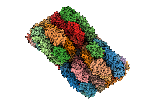
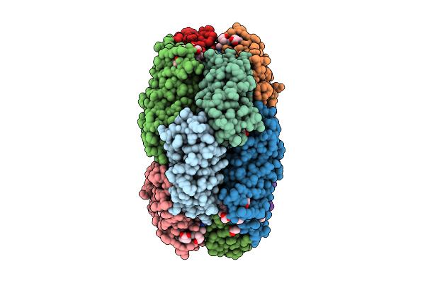
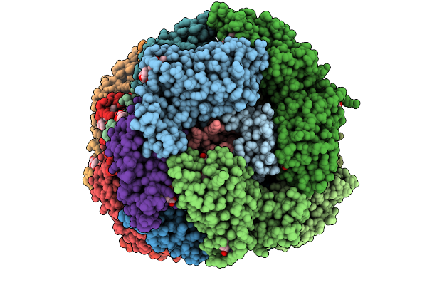
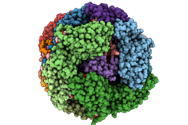
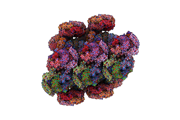
Phycobilisome Allophycocyanin Hexamer C From Gloeobacter Violaceus Pcc 7421
Organism: Gloeobacter violaceus pcc 7421
Method: ELECTRON MICROSCOPY Release Date: 2025-09-24 Classification: PHOTOSYNTHESIS Ligands: CYC |
 |
Organism: Gloeobacter violaceus pcc 7421
Method: ELECTRON MICROSCOPY Release Date: 2025-09-24 Classification: PHOTOSYNTHESIS Ligands: CYC |
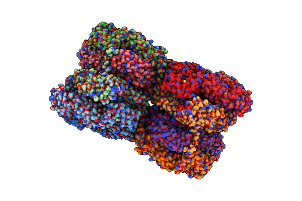 |
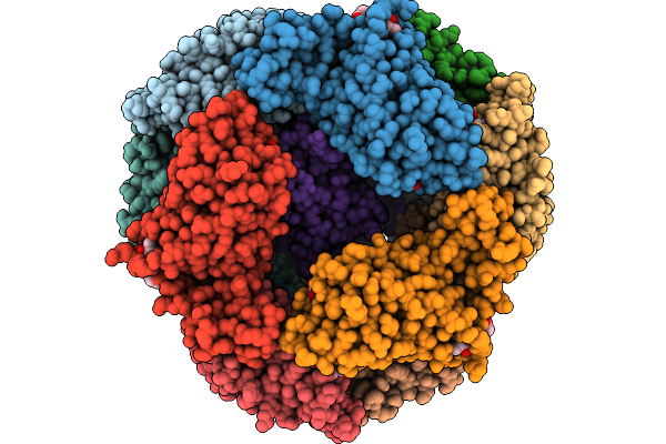
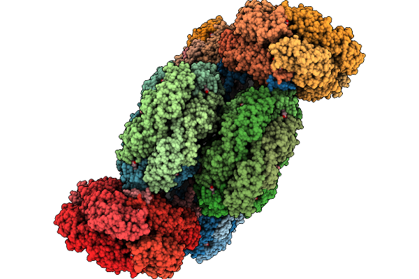
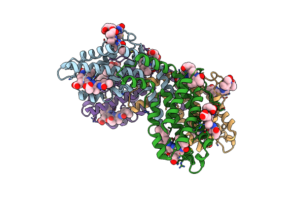
Structure Of The Bicylindrical Allophycocyanin Core Expressed During Far-Red Light Photoacclimation (Farlip)
Organism: Chroococcidiopsis thermalis pcc 7203
Method: ELECTRON MICROSCOPY Release Date: 2025-07-02 Classification: PHOTOSYNTHESIS Ligands: CYC |
 |
Organism: Glycine max
Method: X-RAY DIFFRACTION Release Date: 2025-06-04 Classification: PLANT PROTEIN Ligands: CYC |
 |
Organism: Oscillatoria sp. n9dm
Method: X-RAY DIFFRACTION Release Date: 2025-04-16 Classification: PHOTOSYNTHESIS Ligands: CYC, GOL |
 |
Organism: Synechocystis
Method: X-RAY DIFFRACTION Resolution:3.70 Å Release Date: 2025-03-12 Classification: FLUORESCENT PROTEIN Ligands: CYC |
 |
Organism: Glycine max
Method: X-RAY DIFFRACTION Release Date: 2025-01-08 Classification: PLANT PROTEIN Ligands: CYC, 1PE, PEG, PGE |
 |
Organism: Glycine max
Method: X-RAY DIFFRACTION Release Date: 2025-01-08 Classification: PLANT PROTEIN Ligands: CYC |
 |
Organism: Glycine max
Method: X-RAY DIFFRACTION Release Date: 2025-01-08 Classification: PLANT PROTEIN Ligands: CYC |
 |
Organism: Ceramium secundatum
Method: X-RAY DIFFRACTION Resolution:2.07 Å Release Date: 2024-09-04 Classification: LUMINESCENT PROTEIN Ligands: CYC, PUB |
 |
Organism: Arthrospira sp. fachb-439
Method: ELECTRON MICROSCOPY Release Date: 2024-07-31 Classification: PHOTOSYNTHESIS Ligands: CYC, OEX, CL, CLA, PL9, SQD, LMG, LMT, BCT, LHG, BCR, DGD, PHO, FE2, HEM, CA |
 |
Organism: Synechococcus sp. ja-2-3b'a(2-13)
Method: X-RAY DIFFRACTION Resolution:2.60 Å Release Date: 2024-05-22 Classification: GENE REGULATION Ligands: CYC, EDO, ACT |