
Search Count: 600
All
Selected
 |
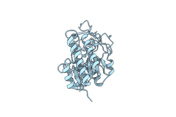
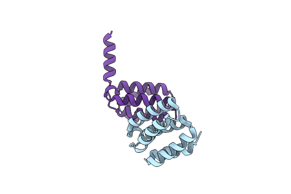
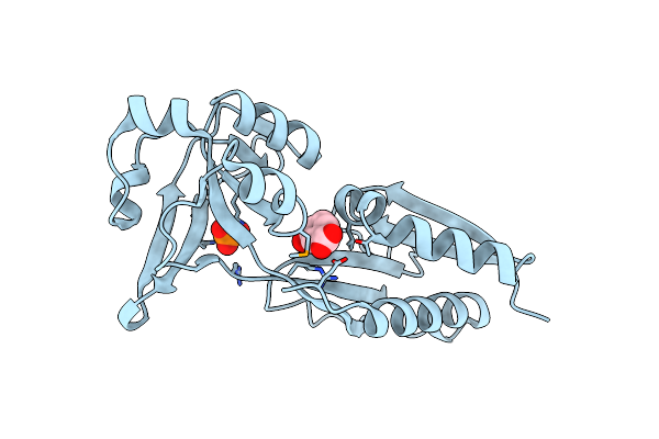
Contact-Dependent Growth Inhibition (Cdi) Immunity Protein From E. Coli O32:H37
Organism: Escherichia coli o32:h37
Method: X-RAY DIFFRACTION Resolution:1.00 Å Release Date: 2023-11-08 Classification: TOXIN Ligands: FE2, NA |
 |
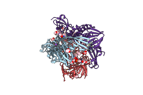
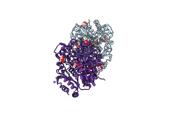
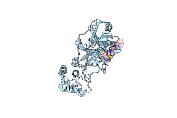
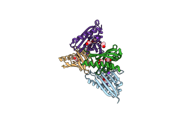
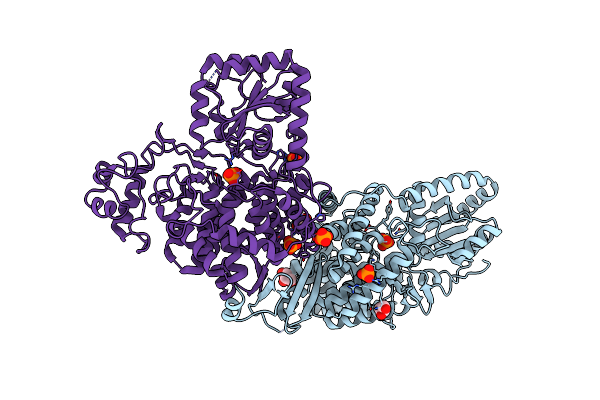
Contact-Dependent Growth Inhibition Toxin-Immunity Protein Complex From E. Coli O32:H37
Organism: Escherichia coli o32:h37
Method: X-RAY DIFFRACTION Resolution:1.83 Å Release Date: 2023-11-08 Classification: TOXIN Ligands: FE2 |
 |
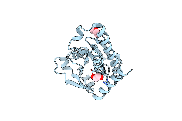
Crystal Structure Of The Dtdp-4-Dehydrorhamnose Reductase From Streptococcus Pneumoniae.
Organism: Streptococcus pneumoniae taiwan19f-14
Method: X-RAY DIFFRACTION Resolution:1.00 Å Release Date: 2023-02-22 Classification: OXIDOREDUCTASE |
 |
Organism: Severe acute respiratory syndrome coronavirus 2
Method: X-RAY DIFFRACTION Resolution:2.17 Å Release Date: 2023-02-22 Classification: HYDROLASE/HYDROLASE INHIBITOR Ligands: ZN, YOO, NA, CL, ACT |
 |
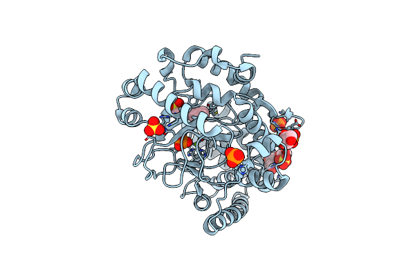
Crystal Structure Of The Threonine Synthase From Streptococcus Pneumoniae In Complex With Pyridoxal 5-Phosphate.
Organism: Streptococcus pneumoniae tigr4
Method: X-RAY DIFFRACTION Resolution:1.43 Å Release Date: 2023-02-15 Classification: LYASE Ligands: CL, NA, PLP, GOL, MLT |
 |
Crystal Structure Of The C-Terminal Fragment Of Aaa Atpase From Streptococcus Pneumoniae.
Organism: Streptococcus pneumoniae tigr4
Method: X-RAY DIFFRACTION Resolution:1.30 Å Release Date: 2023-02-15 Classification: HYDROLASE Ligands: CL |
 |
Crystal Structure Of The Hypothetical Protein (Acx60_00475) From Acinetobacter Baumannii
Organism: Acinetobacter baumannii
Method: X-RAY DIFFRACTION Resolution:1.65 Å Release Date: 2022-11-30 Classification: UNKNOWN FUNCTION Ligands: PEG, EDO |
 |
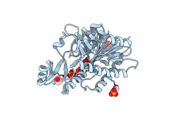
Crystal Structure Of The Catalytic Domain Of The Inosine Monophosphate Dehydrogenase From Listeria Monocytogenes In The Complex With Imp
Organism: Listeria monocytogenes egd-e
Method: X-RAY DIFFRACTION Resolution:2.50 Å Release Date: 2022-09-07 Classification: OXIDOREDUCTASE Ligands: IMP, GOL, FMT |
 |
Organism: Pseudomonas aeruginosa pao1
Method: X-RAY DIFFRACTION Resolution:1.60 Å Release Date: 2022-08-10 Classification: TRANSFERASE Ligands: EDO, FMT, OO9, SO4 |
 |
Thiol-Disulfide Oxidoreductase Tsda, C129S Mutant, From Corynebacterium Diphtheriae
Organism: Corynebacterium diphtheriae
Method: X-RAY DIFFRACTION Resolution:1.10 Å Release Date: 2022-04-20 Classification: OXIDOREDUCTASE Ligands: PGE, EDO, CL |
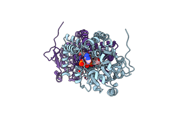 |
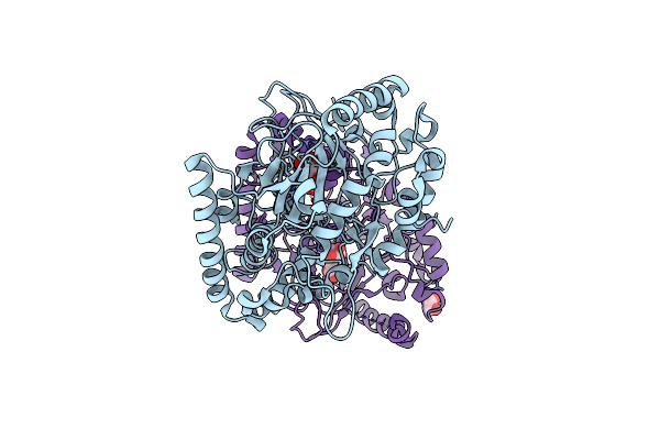
Crystal Structure Of The Mitochondrial Ketol-Acid Reductoisomerase Ilvc From Candida Auris
Organism: [candida] auris
Method: X-RAY DIFFRACTION Resolution:2.43 Å Release Date: 2022-02-16 Classification: ISOMERASE Ligands: NDP, MLI, ACY, MG |
 |
The Crystal Structure Of Sars-Cov-2 Omicron Mpro (P132H) In Complex With Masitinib
Organism: Severe acute respiratory syndrome coronavirus 2
Method: X-RAY DIFFRACTION Resolution:2.09 Å Release Date: 2022-02-16 Classification: HYDROLASE/INHIBITOR Ligands: G65 |
 |
Crystal Structure Of The Putative Oxidoreductase Of Duf1479-Containing Protein Family Ypo2976 From Yersinia Pestis Bound To Caps
Organism: Yersinia pestis co92
Method: X-RAY DIFFRACTION Resolution:1.85 Å Release Date: 2022-02-16 Classification: UNKNOWN FUNCTION Ligands: CXS, MG, PO4, SO4, GOL |
 |
Crystal Structure Of The Putative Oxidoreductase Of Duf1479-Containing Protein Family Ypo2976 From Yersinia Pestis Bound To 2-Oxo-Glutaric Acid
Organism: Yersinia pestis co92
Method: X-RAY DIFFRACTION Resolution:2.41 Å Release Date: 2022-02-16 Classification: UNKNOWN FUNCTION Ligands: MN, AKG, EDO |
 |
Crystal Structure Of The Dna-Binding Domain Of The Lysr Family Transcriptional Regulator Yfba From Yersinia Pestis
Organism: Yersinia pestis co92
Method: X-RAY DIFFRACTION Resolution:1.80 Å Release Date: 2022-02-09 Classification: TRANSCRIPTION Ligands: SO4, CL, GOL, FMT |
 |
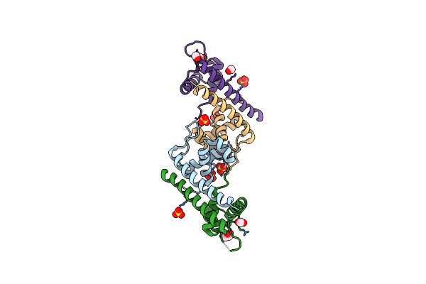
Crystal Structure Of The C-Terminal Ligand-Binding Domain Of The Lysr Family Transcriptional Regulator Yfba From Yersinia Pestis
Organism: Yersinia pestis co92
Method: X-RAY DIFFRACTION Resolution:2.28 Å Release Date: 2022-02-09 Classification: TRANSCRIPTION Ligands: 3HB, PO4 |
 |
Crystal Structure Of The Protein Of Unknown Function Ypo0625 From Yersinia Pestis
Organism: Yersinia pestis co92
Method: X-RAY DIFFRACTION Resolution:2.55 Å Release Date: 2022-02-02 Classification: UNKNOWN FUNCTION Ligands: PEG, K, CL, EDO, GOL |
 |
Crystal Structure Of The Soluble Domain Of The Putative Ompa -Family Membrane Protein Ypo0514 From Yersinia Pestis
Organism: Yersinia pestis co92
Method: X-RAY DIFFRACTION Resolution:2.20 Å Release Date: 2022-01-26 Classification: TRANSPORT PROTEIN Ligands: CA, PO4 |
 |
Crystal Structure Of The Putative Fluoride Ion Transporter Crcb Bab1_1389 From Brucella Abortus
Organism: Brucella abortus 2308
Method: X-RAY DIFFRACTION Resolution:2.10 Å Release Date: 2022-01-26 Classification: UNKNOWN FUNCTION Ligands: EDO, FMT, F, ACT, PEG |
 |
Crystal Structure Of The Thioredox_Dsbh Domain-Containing Uncharacterized Protein Bab1_2064 From Brucella Abortus
Organism: Brucella abortus 2308
Method: X-RAY DIFFRACTION Resolution:2.80 Å Release Date: 2022-01-26 Classification: UNKNOWN FUNCTION Ligands: GOL, PO4, CL |