
Search Count: 48
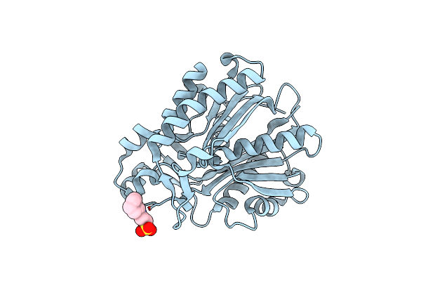 |
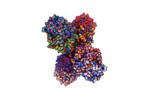
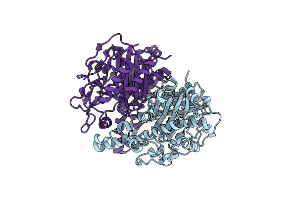
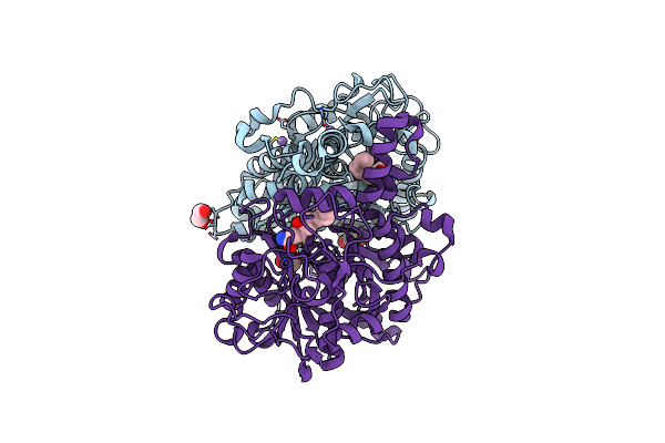
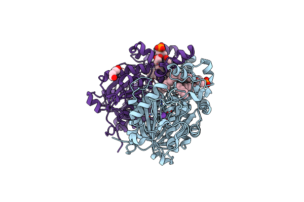
Crystal Structure Of Bacillus Subtilis Fabha, Beta-Ketoacyl Carrier Protein Synthase Iii
Organism: Bacillus subtilis
Method: X-RAY DIFFRACTION Resolution:1.85 Å Release Date: 2024-02-14 Classification: TRANSFERASE Ligands: NHE |
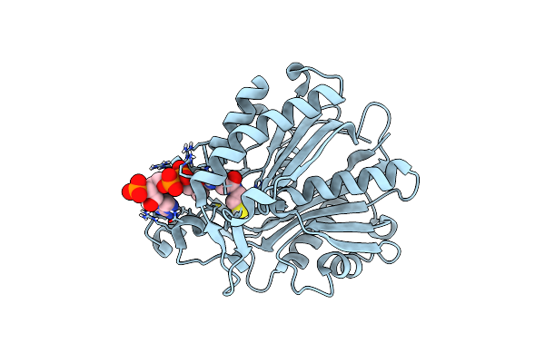 |
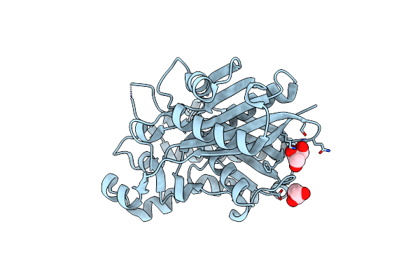
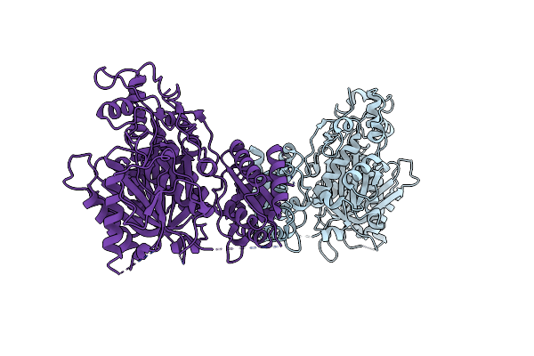
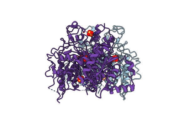
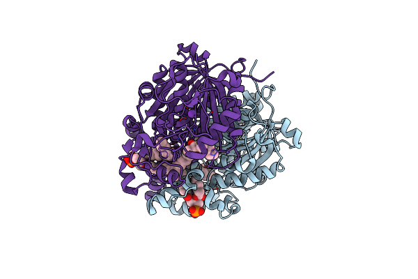
Organism: Bacillus subtilis
Method: X-RAY DIFFRACTION Resolution:2.02 Å Release Date: 2024-02-14 Classification: TRANSFERASE Ligands: COA |
 |
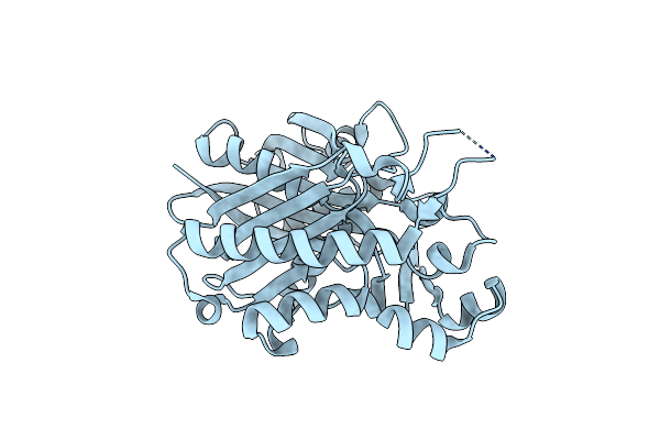
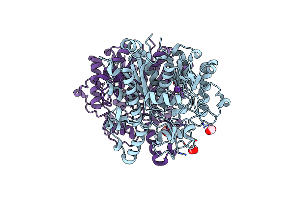
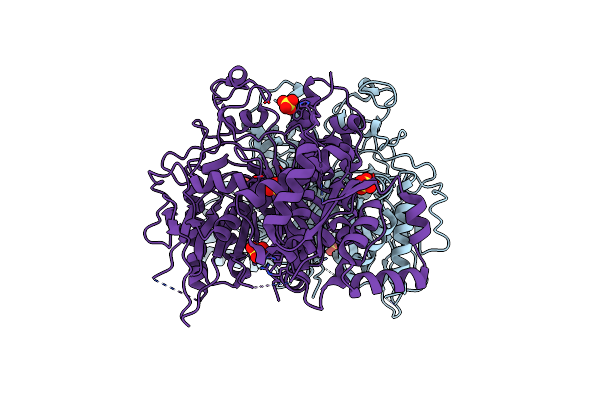
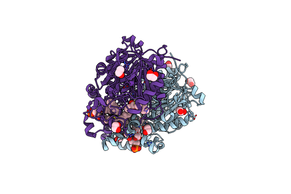
Crystal Structure Of Bacillus Subtilis Fabhb, Beta-Ketoacyl Carrier Protein Synthase Iii
Organism: Bacillus subtilis
Method: X-RAY DIFFRACTION Resolution:2.40 Å Release Date: 2024-02-14 Classification: TRANSFERASE Ligands: GOL |
 |
1.35 Angstrom Resolution Crystal Structure Of Beta-Ketoacyl-Acp Synthase Ii (Fabf) From Listeria Monocytogenes
Organism: Listeria monocytogenes atcc 19115
Method: X-RAY DIFFRACTION Resolution:1.35 Å Release Date: 2016-11-02 Classification: TRANSFERASE |
 |
Organism: Verrucosispora maris (strain ab-18-032)
Method: X-RAY DIFFRACTION Resolution:1.80 Å Release Date: 2016-06-29 Classification: TRANSFERASE |
 |
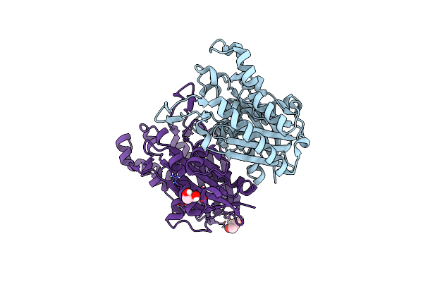
Structural And Functional Characterization Of Pqsbc, A Condensing Enzyme In The Biosynthesis Of The Pseudomonas Aeruginosa Quinolone Signal
Organism: Pseudomonas aeruginosa
Method: X-RAY DIFFRACTION Resolution:2.04 Å Release Date: 2016-02-03 Classification: TRANSFERASE Ligands: PO4, GOL, MPO, TRS, CL, PGE, PEG, NA |
 |
Crystal Structure Of Beta-Ketoacyl-Acp Synthase Iii (Fabh) From Yersinia Pestis With Acetylated Active Site Cysteine
Organism: Yersinia pestis
Method: X-RAY DIFFRACTION Resolution:1.80 Å Release Date: 2015-05-20 Classification: TRANSFERASE Ligands: GOL |
 |
Crystal Structure Of Beta-Ketoacyl-Acp Synthase Iii (Fabh) From Yersinia Pestis
Organism: Yersinia pestis
Method: X-RAY DIFFRACTION Resolution:2.20 Å Release Date: 2015-04-08 Classification: TRANSFERASE |
 |
Crystal Structure Of Beta-Ketoacyl-Acp Synthase Iii (Fabh) From Neisseria Meningitidis
Organism: Neisseria meningitidis fam18
Method: X-RAY DIFFRACTION Resolution:2.45 Å Release Date: 2014-12-31 Classification: TRANSFERASE Ligands: GOL |
 |
Crystal Structure Of Beta-Ketoacyl-Acp Synthase Ii (Fabf) From Yersinia Pestis
Organism: Yersinia pestis
Method: X-RAY DIFFRACTION Resolution:2.70 Å Release Date: 2014-12-17 Classification: TRANSFERASE |
 |
The Structure Of Beta-Ketoacyl -(Acyl Carrier Protein) Synthase Ii (Fabf) From Neisseria Meningitidis
Organism: Neisseria meningitidis
Method: X-RAY DIFFRACTION Resolution:2.10 Å Release Date: 2014-06-04 Classification: TRANSFERASE Ligands: K, FMT |
 |
Crystal Structure Of Beta-Ketoacyl-Acp Synthase Ii (Fabf) From Bacillus Subtilis
Organism: Bacillus subtilis subsp. subtilis
Method: X-RAY DIFFRACTION Resolution:1.80 Å Release Date: 2014-04-02 Classification: TRANSFERASE Ligands: K, GOL |
 |
Crystal Structure Of Bacillus Subtilis Beta-Ketoacyl-Acp Synthase Ii (Fabf) In A Non-Covalent Complex With Cerulenin
Organism: Bacillus subtilis subsp. subtilis
Method: X-RAY DIFFRACTION Resolution:1.67 Å Release Date: 2014-04-02 Classification: transferase/transferase inhibitor Ligands: K, GOL, 1X9 |
 |
Crystal Structure Of Bacillus Subtilis Beta-Ketoacyl-Acp Synthase Ii (Fabf) In A Covalent Complex With Cerulenin
Organism: Bacillus subtilis subsp. subtilis
Method: X-RAY DIFFRACTION Resolution:2.10 Å Release Date: 2014-04-02 Classification: TRANSFERASE Ligands: 1XG, NA, CL, EDO |
 |
Crystal Structure Of The Second Ketosynthase From The Bacillaene Polyketide Synthase
Organism: Bacillus subtilis subsp. subtilis
Method: X-RAY DIFFRACTION Resolution:1.95 Å Release Date: 2014-02-19 Classification: TRANSFERASE Ligands: SO4 |
 |
Crystal Structure Of The Second Ketosynthase From The Bacillaene Polyketide Synthase Bound To Its Natural Intermediate
Organism: Bacillus subtilis subsp. subtilis
Method: X-RAY DIFFRACTION Resolution:2.30 Å Release Date: 2014-02-19 Classification: TRANSFERASE |
 |
Crystal Structure Of The Second Ketosynthase From The Bacillaene Polyketide Synthase Bound To A Hexanoyl Substrate Mimic
Organism: Bacillus subtilis subsp. subtilis
Method: X-RAY DIFFRACTION Resolution:2.89 Å Release Date: 2014-02-19 Classification: TRANSFERASE Ligands: SO4 |
 |
Organism: Mycobacterium tuberculosis h37rv
Method: X-RAY DIFFRACTION Resolution:1.70 Å Release Date: 2013-10-09 Classification: TRANSFERASE Ligands: K, M7U, EPE, PEG, NA |
 |
Crystal Structure Of M. Tuberculosis C171Q Kasa In Complex With Thiolactomycin (Tlm)
Organism: Mycobacterium tuberculosis h37rv
Method: X-RAY DIFFRACTION Resolution:1.95 Å Release Date: 2013-10-09 Classification: TRANSFERASE Ligands: EDO, K, TLM, M7U, EPE |
 |
Organism: Mycobacterium tuberculosis
Method: X-RAY DIFFRACTION Resolution:1.80 Å Release Date: 2013-10-09 Classification: TRANSFERASE Ligands: EDO, K, TLE, M7U |