
Search Count: 145
 |
Organism: Ostrinia furnacalis
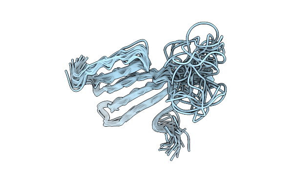
Method: SOLID-STATE NMR Release Date: 2025-10-22 Classification: STRUCTURAL PROTEIN |
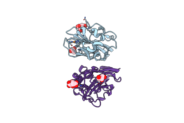 |
Organism: Serratia marcescens
Method: X-RAY DIFFRACTION Resolution:1.46 Å Release Date: 2024-06-12 Classification: OXIDOREDUCTASE Ligands: CU, GOL, CIT |
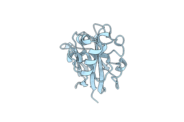 |
Sub-Atomic Resolution Structure Of The Chitin-Binding Protein D (Cbpd) From Pseudomonas Aeruginosa
Organism: Pseudomonas aeruginosa pa14
Method: X-RAY DIFFRACTION Resolution:0.75 Å Release Date: 2023-06-28 Classification: OXIDOREDUCTASE Ligands: CL |
 |
Crystal Structure Of The Cerato-Platanin-Like Protein Cpl1 From Ustilago Maydis
Organism: Ustilago maydis 521
Method: X-RAY DIFFRACTION Resolution:1.90 Å Release Date: 2023-05-17 Classification: SUGAR BINDING PROTEIN |
 |
Organism: Ustilago hordei
Method: X-RAY DIFFRACTION Resolution:1.35 Å Release Date: 2023-05-17 Classification: SUGAR BINDING PROTEIN |
 |
Organism: Thermothelomyces thermophilus
Method: X-RAY DIFFRACTION Resolution:2.65 Å Release Date: 2023-03-15 Classification: METAL BINDING PROTEIN Ligands: NAG, CU, ACT |
 |
Organism: Cucumis sativus
Method: X-RAY DIFFRACTION Resolution:1.20 Å Release Date: 2023-03-08 Classification: SUGAR BINDING PROTEIN Ligands: NBZ, EDO |
 |
Organism: Cucumis sativus
Method: X-RAY DIFFRACTION Resolution:1.90 Å Release Date: 2023-03-08 Classification: SUGAR BINDING PROTEIN Ligands: EDO |
 |
Organism: Cucumis sativus
Method: X-RAY DIFFRACTION Resolution:2.50 Å Release Date: 2023-03-08 Classification: SUGAR BINDING PROTEIN |
 |
Organism: Cucumis sativus
Method: X-RAY DIFFRACTION Resolution:1.85 Å Release Date: 2023-03-08 Classification: SUGAR BINDING PROTEIN |
 |
Organism: Cucumis sativus
Method: X-RAY DIFFRACTION Resolution:2.50 Å Release Date: 2023-02-01 Classification: SUGAR BINDING PROTEIN Ligands: PT |
 |
Crystal Structure Of Pseudomonas Aeruginosa Lytic Polysaccharide Monooxygenase Cbpd
Organism: Pseudomonas aeruginosa (strain atcc 15692 / dsm 22644 / cip 104116 / jcm 14847 / lmg 12228 / 1c / prs 101 / pao1)
Method: X-RAY DIFFRACTION Resolution:3.00 Å Release Date: 2022-07-20 Classification: OXIDOREDUCTASE Ligands: NH4 |
 |
Crystal Structure Of A Chitin-Binding Protein From Moringa Oleifera Seeds (Mo-Cbp4)
Organism: Moringa oleifera
Method: X-RAY DIFFRACTION Resolution:1.90 Å Release Date: 2021-01-27 Classification: PLANT PROTEIN Ligands: ACT, CL |
 |
Organism: Photorhabdus laumondii subsp. laumondii (strain dsm 15139 / cip 105565 / tt01)
Method: X-RAY DIFFRACTION Resolution:1.60 Å Release Date: 2020-01-15 Classification: OXIDOREDUCTASE Ligands: CU |
 |
Organism: Chenopodium quinoa
Method: SOLUTION NMR Release Date: 2019-05-15 Classification: PLANT PROTEIN |
 |
Crystal Structure Of Aa10 Lytic Polysaccharide Monooxygenase From Tectaria Macrodonta
Organism: Tectaria macrodonta
Method: X-RAY DIFFRACTION Resolution:2.20 Å Release Date: 2019-04-24 Classification: SUGAR BINDING PROTEIN Ligands: CU, SO4, GOL, MG |
 |
Organism: Micromonospora aurantiaca atcc 27029
Method: X-RAY DIFFRACTION Resolution:1.08 Å Release Date: 2017-12-13 Classification: CARBOHYDRATE Ligands: CU |
 |
Organism: Listeria monocytogenes serotype 1/2a (strain 10403s)
Method: X-RAY DIFFRACTION Resolution:1.10 Å Release Date: 2017-08-09 Classification: CHITIN-BINDING PROTEIN Ligands: CU, P33 |
 |
Organism: Flavobacterium johnsoniae (strain atcc 17061 / dsm 2064 / uw101)
Method: X-RAY DIFFRACTION Resolution:2.30 Å Release Date: 2017-02-08 Classification: CHITIN-BINDING PROTEIN |
 |
Structure Of Fjoh_4558, A Chitin-Binding Susd Homolog From Flavobacterium Johnsoniae
Organism: Flavobacterium johnsoniae
Method: X-RAY DIFFRACTION Resolution:1.39 Å Release Date: 2017-02-08 Classification: Chitin-binding protein Ligands: EDO |