
Search Count: 4
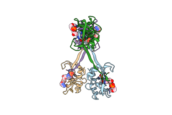 |
X-Ray Structure Of The C-Terminal Swapped Dimer Of P114A Variant Of Ribonuclease A
Organism: Bos taurus
Method: X-RAY DIFFRACTION Resolution:2.18 Å Release Date: 2012-02-15 Classification: HYDROLASE Ligands: CGP, PO4 |
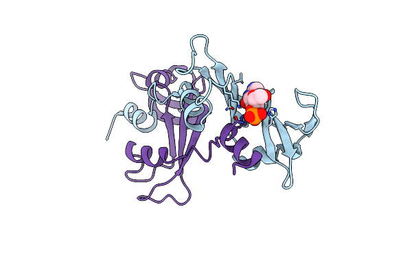 |
X-Ray Structure Of The Non Covalent Swapped Form Of The Q28L/K31C/S32C Mutant Of Bovine Pancreatic Ribonuclease In Complex With 2'-Deoxycytidine-2'-Deoxyguanosine-3',5'-Monophosphate
Organism: Bos taurus
Method: X-RAY DIFFRACTION Resolution:1.94 Å Release Date: 2009-03-24 Classification: HYDROLASE Ligands: CGP |
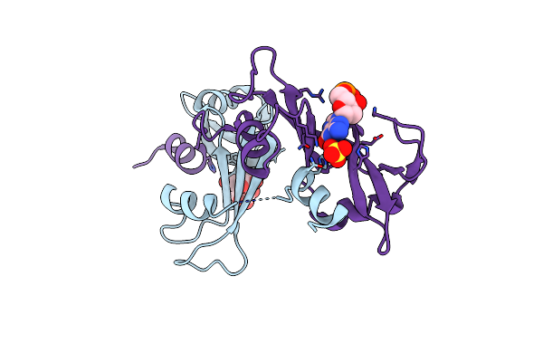 |
X-Ray Structure Of The Non Covalent Swapped Form Of The A19P/Q28L/K31C/S32C Mutant Of Bovine Pancreatic Ribonuclease In Complex With 2'-Deoxycytidine-2'-Deoxyguanosine-3',5'-Monophosphate
Organism: Bos taurus
Method: X-RAY DIFFRACTION Resolution:1.90 Å Release Date: 2009-03-24 Classification: HYDROLASE Ligands: CGP, SO4 |
 |
Structure Of The Crystalline Complex Of Deoxycytidylyl-3',5'-Guanosine (3',5'-Dcpdg) Co-Crystalised With Ribonuclease At 1.9 Angstroms Resolution. Retrobinding In Pancreatic Rnasea Is Independent Of Mode Of Inhibitor Intromission
Organism: Bos taurus
Method: X-RAY DIFFRACTION Resolution:1.90 Å Release Date: 1995-09-15 Classification: HYDROLASE (ENDORIBONUCLEASE) Ligands: PO4, CGP |