
Search Count: 3,380
All
Selected
 |
Organism: Homo sapiens
Method: X-RAY DIFFRACTION Resolution:2.85 Å Release Date: 2026-03-04 Classification: CELL CYCLE Ligands: A1B7H, CIT |
 |
Organism: Homo sapiens
Method: X-RAY DIFFRACTION Resolution:1.60 Å Release Date: 2026-02-25 Classification: CELL CYCLE Ligands: PO4, GOL |
 |
Organism: Homo sapiens
Method: ELECTRON MICROSCOPY Release Date: 2026-02-18 Classification: TRANSLATION Ligands: KGN |
 |
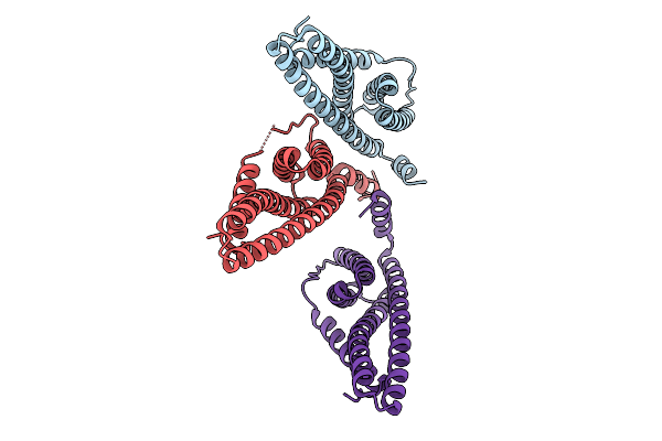
Organism: Homo sapiens
Method: ELECTRON MICROSCOPY Release Date: 2026-02-18 Classification: CELL CYCLE Ligands: ATP, ADP |
 |
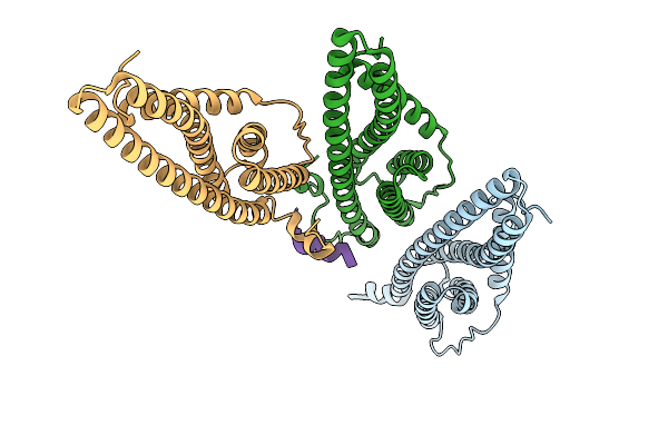
Organism: Homo sapiens
Method: ELECTRON MICROSCOPY Release Date: 2026-02-18 Classification: CELL CYCLE Ligands: ATP, ADP |
 |
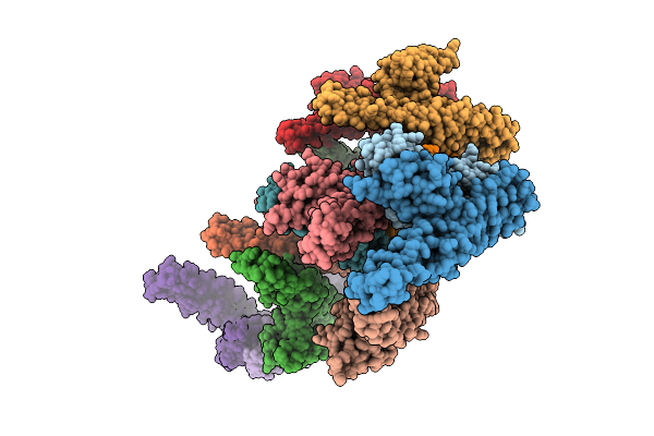
Organism: Homo sapiens
Method: ELECTRON MICROSCOPY Release Date: 2026-02-18 Classification: CELL CYCLE Ligands: ATP, ADP |
 |
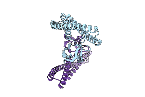
Organism: Homo sapiens
Method: ELECTRON MICROSCOPY Release Date: 2026-02-18 Classification: CELL CYCLE |
 |
Organism: Homo sapiens
Method: ELECTRON MICROSCOPY Resolution:3.80 Å Release Date: 2026-02-11 Classification: CELL CYCLE |
 |
Cryo-Em Structure Of The Sah Domain Of Caenorhabditis Elegans Icp-1 Bound To The Paclitaxel-Stabilized Microtubule
Organism: Caenorhabditis elegans, Sus scrofa
Method: ELECTRON MICROSCOPY Release Date: 2026-02-04 Classification: CELL CYCLE Ligands: GTP, ZN, MG, GDP, TA1 |
 |
Cryo-Em Structure Of The Sah Domain Of Human Incenp Bound To The Paclitaxel-Stabilized Microtubule
Organism: Homo sapiens, Sus scrofa
Method: ELECTRON MICROSCOPY Release Date: 2026-02-04 Classification: CELL CYCLE Ligands: GTP, ZN, MG, GDP, TA1 |
 |
Cryo-Em Structure Of The Human Hec1-Nuf2 Dimer Bound To The Paclitaxel-Stabilized Microtubule
Organism: Homo sapiens, Sus scrofa
Method: ELECTRON MICROSCOPY Release Date: 2026-02-04 Classification: CELL CYCLE Ligands: GTP, MG, GDP, TA1 |
 |
Cryo-Em Structure Of The Xenopus Laevis Mitotic Centromere-Associated Kinesin (Mcak) Bound To The Paclitaxel-Stabilized Microtubule
Organism: Xenopus laevis, Sus scrofa
Method: ELECTRON MICROSCOPY Release Date: 2026-02-04 Classification: CELL CYCLE Ligands: GTP, MG, GDP, TA1 |
 |
Cryo-Em Structure Of The Xenopus Laevis Mitotic Centromere-Associated Kinesin (Mcak) Bound To The Paclitaxel-Stabilized Microtubule
Organism: Xenopus laevis, Sus scrofa
Method: ELECTRON MICROSCOPY Release Date: 2026-02-04 Classification: CELL CYCLE Ligands: GTP, MG, GDP, TA1 |
 |
Organism: Homo sapiens
Method: ELECTRON MICROSCOPY Release Date: 2026-02-04 Classification: CELL CYCLE Ligands: A1B7G, ZN |
 |
Cryo-Em Structure Of The Xenopus Laevis Mitotic Centromere-Associated Kinesin (Mcak) Bound To The Paclitaxel-Stabilized Microtubule
Organism: Xenopus laevis, Sus scrofa
Method: ELECTRON MICROSCOPY Release Date: 2026-02-04 Classification: CELL CYCLE Ligands: MG, GTP, GDP, TA1 |
 |
Organism: Sulfolobus acidocaldarius
Method: ELECTRON MICROSCOPY Release Date: 2026-01-28 Classification: CELL CYCLE |
 |
Organism: Sulfolobus acidocaldarius
Method: ELECTRON MICROSCOPY Release Date: 2026-01-28 Classification: CELL CYCLE |
 |
Organism: Sulfolobus acidocaldarius
Method: ELECTRON MICROSCOPY Release Date: 2026-01-28 Classification: CELL CYCLE |
 |
Organism: Saccharolobus islandicus
Method: X-RAY DIFFRACTION Resolution:2.85 Å Release Date: 2026-01-28 Classification: CELL CYCLE |
 |
Organism: Saccharolobus islandicus
Method: ELECTRON MICROSCOPY Release Date: 2026-01-28 Classification: CELL CYCLE |