
Search Count: 66
 |
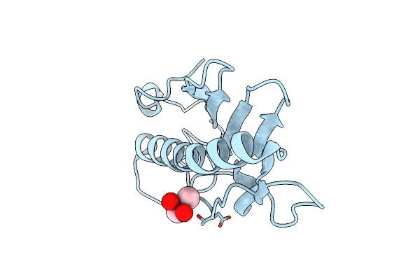
Organism: Homo sapiens
Method: X-RAY DIFFRACTION Resolution:2.00 Å Release Date: 2025-05-28 Classification: IMMUNE SYSTEM Ligands: CAD, EDO, PEG, PGE |
 |
Organism: Pseudomonas aeruginosa
Method: X-RAY DIFFRACTION Resolution:1.30 Å Release Date: 2024-12-11 Classification: CELL ADHESION Ligands: CAD |
 |
Organism: Homo sapiens
Method: X-RAY DIFFRACTION Resolution:2.94 Å Release Date: 2024-04-10 Classification: TRANSCRIPTION Ligands: CAD, GOL, YF8 |
 |
Organism: Pseudomonas aeruginosa
Method: X-RAY DIFFRACTION Resolution:2.10 Å Release Date: 2023-09-27 Classification: CYTOSOLIC PROTEIN Ligands: PPI, GOL, CAD, GLY, 1PE, PG4 |
 |
Organism: Synthetic construct
Method: X-RAY DIFFRACTION Resolution:2.01 Å Release Date: 2023-08-09 Classification: DNA Ligands: CAD |
 |
Organism: Homo sapiens
Method: X-RAY DIFFRACTION Resolution:2.90 Å Release Date: 2023-05-31 Classification: RNA Ligands: K, CAD |
 |
Organism: Homo sapiens
Method: X-RAY DIFFRACTION Resolution:1.96 Å Release Date: 2022-03-16 Classification: HYDROLASE/Immune System Ligands: Z07, GDP, MG, ACT, CAD, P15, EDO, DMS |
 |
Crystal Structure Of L182E D-Succinylase (Dsa) From Cupriavidus Sp. P4-10-C
Organism: Cupriavidus sp. p4-10-c
Method: X-RAY DIFFRACTION Resolution:1.95 Å Release Date: 2022-01-26 Classification: HYDROLASE Ligands: CAD, GOL, SIN, CO3, CA, CL |
 |
Crystal Structure Of A Thermostable Green Fluorescent Protein (Tgp) With A Synthetic Nanobody (Sb92)
Organism: Galaxea fascicularis, Synthetic construct
Method: X-RAY DIFFRACTION Resolution:2.77 Å Release Date: 2021-09-08 Classification: FLUORESCENT PROTEIN Ligands: GOL, CAD, ACT, CAC |
 |
Crystal Structure Of A Biodegradable Plastic-Degrading Cutinase From Paraphoma Sp. B47-9.
Organism: Paraphoma sp. b47-9
Method: X-RAY DIFFRACTION Resolution:1.27 Å Release Date: 2020-09-30 Classification: HYDROLASE Ligands: CAD, NA |
 |
Crystal Structure Of A Biodegradable Plastic-Degrading Cutinase From Paraphoma Sp. B47-9 Solved By Getting The Phase From Anomalous Scattering Of Uncovalently Coordinated Arsenic (Cacodylate).
Organism: Paraphoma sp. b47-9
Method: X-RAY DIFFRACTION Resolution:1.36 Å Release Date: 2020-09-30 Classification: HYDROLASE Ligands: CAD, NA |
 |
Crystal Structure Of A Biodegradable Plastic-Degrading Cutinase-Like Enzyme From The Phyllosphere Yeast, Pseudozyma Antarctica, Solved By Getting The Phase From Anomalous Scattering Of Uncovalently Coordinated Arsenic (Cacodylate).
Organism: Pseudozyma antarctica
Method: X-RAY DIFFRACTION Resolution:1.70 Å Release Date: 2020-09-16 Classification: HYDROLASE Ligands: CAD, NA |
 |
Organism: Homo sapiens
Method: X-RAY DIFFRACTION Resolution:1.69 Å Release Date: 2018-01-17 Classification: APOPTOSIS Ligands: CAD |
 |
Organism: Mycobacterium tuberculosis
Method: X-RAY DIFFRACTION Resolution:2.04 Å Release Date: 2017-10-11 Classification: TRANSFERASE Ligands: ACO, GLU, CAD |
 |
Structure Of Bovine Endothelial Nitric Oxide Synthase Heme Domain In Complex With 4-(2-(((2-Aminoquinolin-7-Yl)Methyl)Amino)Ethyl)-2-Methylbenzonitrile
Organism: Bos taurus
Method: X-RAY DIFFRACTION Resolution:2.15 Å Release Date: 2017-08-16 Classification: OXIDOREDUCTASE/OXIDOREDUCTASE INHIBITOR Ligands: HEM, H4B, 9P7, ACT, GOL, ZN, CAD |
 |
Structural Studies Of A Thermophilic Esterase From Thermogutta Terrifontis (L37A Mutant With Butyrate Bound)
Organism: Planctomycetes bacterium r1
Method: X-RAY DIFFRACTION Resolution:1.08 Å Release Date: 2015-06-10 Classification: HYDROLASE Ligands: CAD, BUA, PGE |
 |
Structural Studies Of A Thermophilic Esterase From Thermogutta Terrifontis (Cacodylate Complex)
Organism: Planctomycetes bacterium r1
Method: X-RAY DIFFRACTION Resolution:1.06 Å Release Date: 2015-06-10 Classification: HYDROLASE Ligands: CAD, PEG, CL |
 |
Crystal Structure Of 2-C-Methyl-D-Erythritol 4-Phosphate Cytidylyltransferase (Ispd) From Burkholderia Thailandensis Complexed With Ctp
Organism: Burkholderia thailandensis
Method: X-RAY DIFFRACTION Resolution:2.30 Å Release Date: 2015-04-29 Classification: TRANSFERASE Ligands: CTP, MG, ACT, CAD, GOL |
 |
Structure Of Bovine Endothelial Nitric Oxide Synthase Heme Domain In Complex With 3,5-Bis(2-(6-Amino-4-Methylpyridin-2-Yl)Ethyl)Benzonitrile
Organism: Bos taurus
Method: X-RAY DIFFRACTION Resolution:2.25 Å Release Date: 2013-04-24 Classification: Oxidoreductase/Oxidoreductase inhibitor Ligands: HEM, H4B, 1EV, ACT, GOL, CAD, ZN |
 |
The Crystal Structure Of Pyrazinamidase/Nicotinamidase From Streptococcus Mutans Ua159
Organism: Streptococcus mutans
Method: X-RAY DIFFRACTION Resolution:1.70 Å Release Date: 2012-05-16 Classification: HYDROLASE Ligands: ZN, CAD |