
Search Count: 11
 |
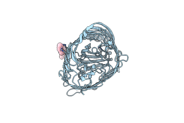
Organism: Xenopsylla cheopis
Method: X-RAY DIFFRACTION Resolution:1.75 Å Release Date: 2024-01-17 Classification: LIPID BINDING PROTEIN Ligands: SRO, SO4, PAM |
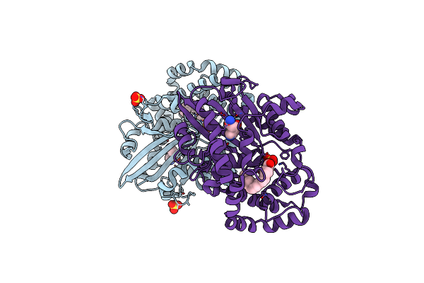
 |
Organism: Xenopsylla cheopis, Homo sapiens
Method: X-RAY DIFFRACTION Resolution:2.15 Å Release Date: 2021-12-22 Classification: BLOOD CLOTTING Ligands: NAG, NA |
 |
Crystal Structure Of Lxrbeta (Nuclear Receptor Subfamily 1, Group H, Member 2) Complexed With Bms-852927
Organism: Homo sapiens
Method: X-RAY DIFFRACTION Resolution:2.40 Å Release Date: 2016-11-02 Classification: TRANSCRIPTION Ligands: 6OX, BU1 |
 |
Crystal Structure Of Nuclear Receptor Subfamily 1, Group H, Member 2 (Lxrb) Complexed With Partial Agonist
Organism: Homo sapiens
Method: X-RAY DIFFRACTION Resolution:2.04 Å Release Date: 2014-12-31 Classification: TRANSCRIPTION/AGONIST Ligands: BU1, 652 |
 |
Crystal Structure Of The C-Terminal Part Of Rhie From Burkholderia Rhizoxinica
Organism: Burkholderia rhizoxinica
Method: X-RAY DIFFRACTION Resolution:2.14 Å Release Date: 2013-09-18 Classification: TRANSFERASE Ligands: GOL |
 |
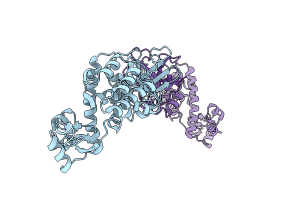
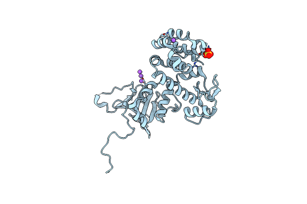
The Crystal Structure Of The Ferric Yersiniabactin Uptake Receptor Fyua From Yersinia Pestis
Organism: Yersinia pestis
Method: X-RAY DIFFRACTION Resolution:3.20 Å Release Date: 2012-06-20 Classification: METAL TRANSPORT Ligands: LDA |
 |
Organism: Yersinia pestis
Method: X-RAY DIFFRACTION Resolution:2.09 Å Release Date: 2012-06-20 Classification: TOXIN |
 |
The Crystal Structure Of Pesticin-T4 Lysozyme Hybrid Stabilized By Engineered Disulfide Bonds
Organism: Yersinia pestis, Enterobacteria phage t4
Method: X-RAY DIFFRACTION Resolution:1.74 Å Release Date: 2012-06-20 Classification: TOXIN, HYDROLASE Ligands: SO4, NA |
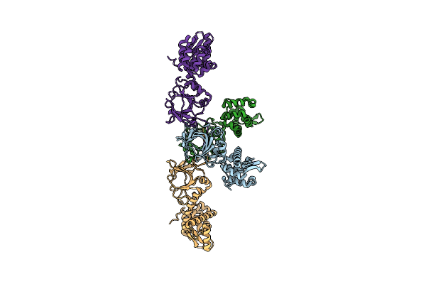 |
The Crystal Structure Of An Engineered Phage Lysin Containing The Binding Domain Of Pesticin And The Killing Domain Of T4-Lysozyme
Organism: Yersinia pestis, Enterobacteria phage t4
Method: X-RAY DIFFRACTION Resolution:2.60 Å Release Date: 2012-06-20 Classification: TOXIN, HYDROLASE |
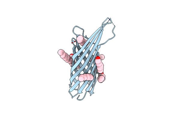 |
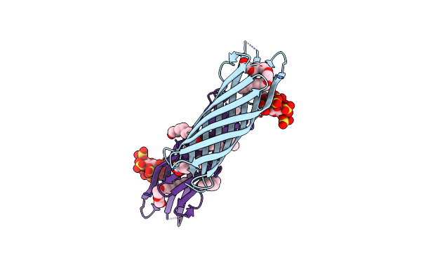
The Crystal Structure Of Ail, The Attachment Invasion Locus Protein Of Yersinia Pestis
Organism: Yersinia pestis
Method: X-RAY DIFFRACTION Resolution:1.80 Å Release Date: 2011-11-23 Classification: CELL INVASION Ligands: C8E |
 |
The Crystal Structure Of Ail, The Attachment Invasion Locus Protein Of Yersinia Pestis, In Complex With The Heparin Analogue Sucrose Octasulfate
Organism: Yersinia pestis
Method: X-RAY DIFFRACTION Resolution:1.85 Å Release Date: 2011-11-23 Classification: CELL INVASION Ligands: C8E |